How to Read NMR Spectra: ¹H NMR (Proton NMR) Interpretation Step by Step
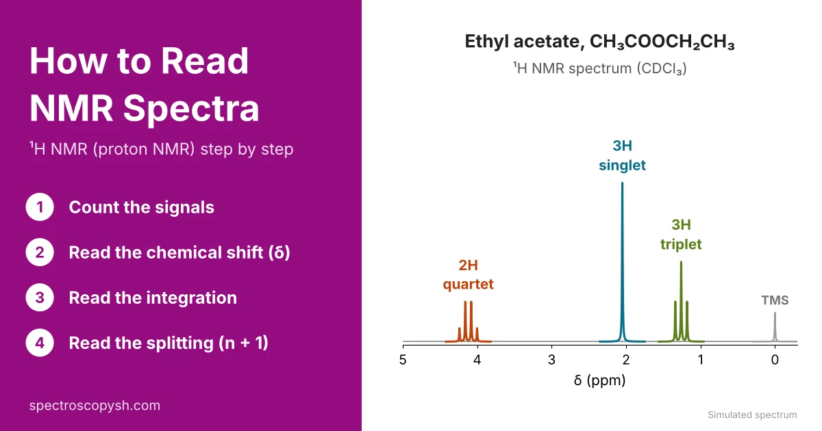
How to read NMR spectra: to read an NMR spectrum, answer four questions: how many signals (kinds of hydrogen), where they appear (chemical shift, δ), how big they are (integration) and how split they are (number of neighbouring protons, n + 1 rule). Each answer gives one piece of the molecule; together they give the structure.
What Information Can Be Obtained from an NMR Spectrum?
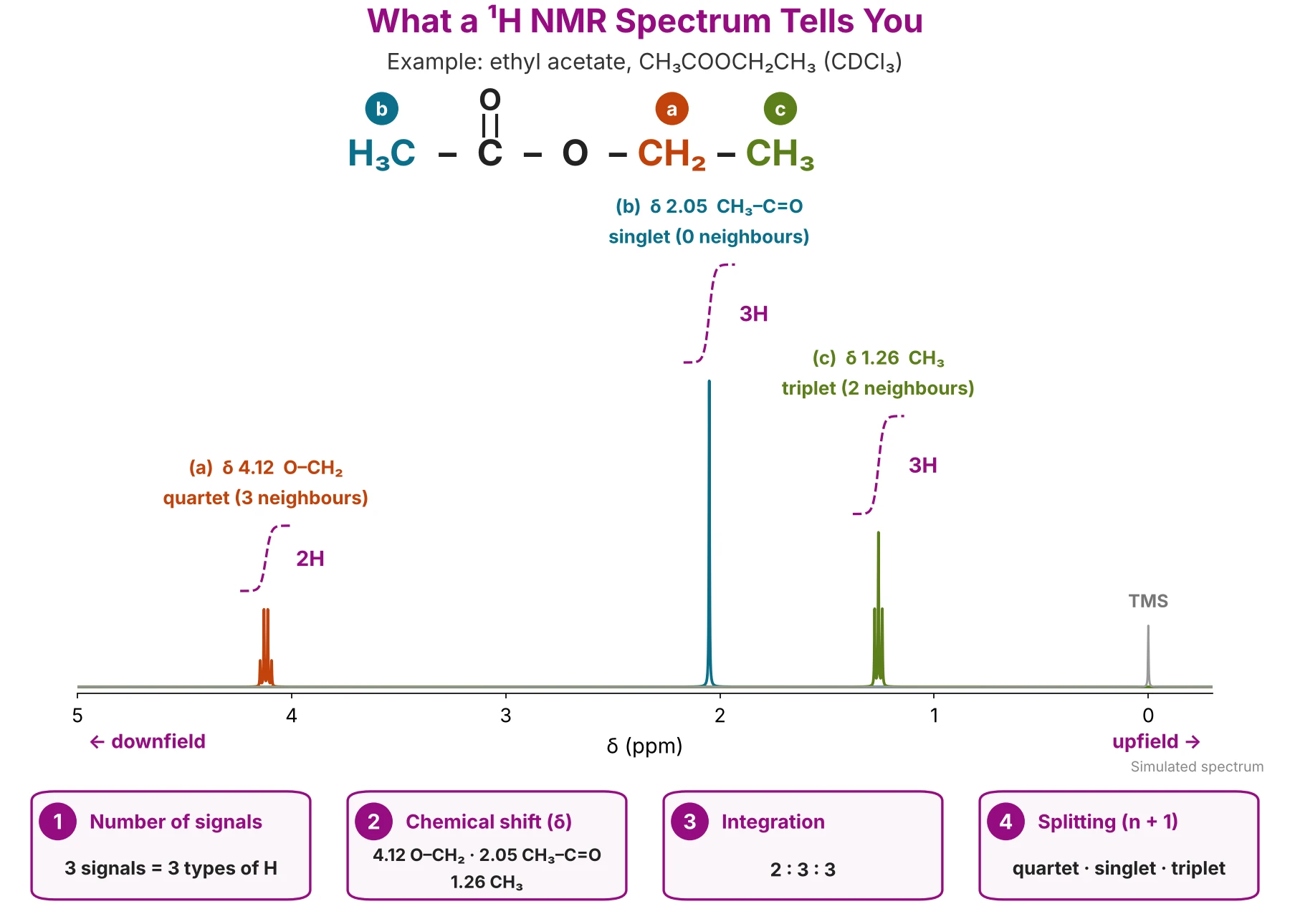
1. Number of Signals
Tells you how many types of protons (chemically different hydrogens) are in the molecule.
3 signals = 3 types of protons; 4 signals = 4 types of protons.
2. Chemical Shift (δ)
Tells you which type of protons are present in the molecule. For example:
- ≈ 2.1 ppm → CH₃ next to C=O (methyl ketone or ester)
- 6.5–8 ppm → aromatic ring (benzene) protons
- 9–10 ppm → aldehyde proton (CHO)
- 10–12 ppm → carboxylic acid proton (COOH)
3. Integration
Tells you the relative number of protons under each peak. NMR integration is the area under each signal, measured by the NMR instrument and shown as a step curve or a number under (or above) the peak.
- Usually it gives a ratio of protons, not the actual number.
- Sometimes the ratio equals the actual number. Example: toluene (methylbenzene), C₆H₅CH₃ shows a 3 : 5 ratio = 3 CH₃ H and 5 aromatic H.
4. Splitting (n + 1 Rule)
Tells you the number of neighbouring protons (on the next carbon) for the proton under study. A proton with n neighbouring protons gives n + 1 peaks:
- Singlet → no neighbouring proton
- Doublet → 1 neighbouring proton
- Triplet → 2 neighbouring protons
- Quartet → 3 neighbouring protons
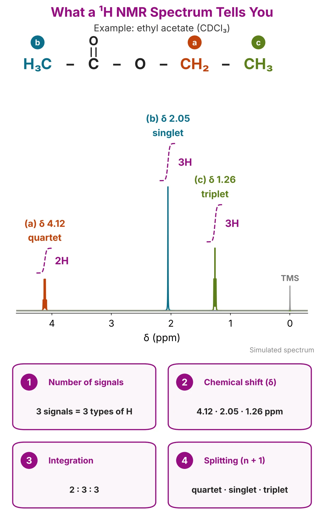
Nuclear magnetic resonance (NMR) spectroscopy records how ¹H or ¹³C nuclei in a magnetic field absorb radio waves. This guide shows how to interpret H NMR and ¹³C nuclear magnetic resonance spectra step by step, so you can read HNMR data for any unknown compound.
Before You Start: Molecular Formula and DBE
Get the molecular formula first (from the mass spectrum), then the degree of unsaturation (DBE):
DBE = C − H/2 − X/2 + N/2 + 1
Example: ethyl acetate, C₄H₈O₂ → DBE = 4 − 4 + 1 = 1 (one C=O). Now read the NMR. See how the formula comes from the mass spectrum in ethyl acetate spectra, Method 1.
What Is an NMR Spectrum?
An NMR spectrum is a graph of signal intensity (y-axis) against chemical shift, δ, in parts per million (ppm) (x-axis). It is recorded by a nuclear magnetic resonance spectrometer, for example at 400 MHz. Each signal (peak) comes from one kind of hydrogen in a ¹H NMR spectrum, or one kind of carbon in a ¹³C NMR spectrum.

x-Axis: Chemical Shift (δ, ppm)
- The ¹H NMR scale runs from 0 to 12 ppm; the ¹³C NMR scale runs from 0 to 220 ppm.
- δ increases from right to left.
- δ (ppm) = shift from TMS (Hz) ÷ spectrometer frequency (MHz). So δ is the same on any instrument: a signal at δ 2.20 is 880 Hz from TMS at 400 MHz and 660 Hz at 300 MHz.
y-Axis: Signal Intensity
- Shows how strong each signal is; usually printed without numbers.
- The area under a signal (integration), not its height, gives the number of hydrogens (see Step 3).
TMS: The Zero Point (δ 0.00)
Tetramethylsilane (TMS) is added as the reference; its single signal is set at δ 0.00 ppm, and every other signal is measured from it.
Downfield and Upfield
- Downfield (left): deshielded protons, large δ, higher frequency.
- Upfield (right): shielded protons, small δ, closer to TMS.
- Why some protons are shielded and others deshielded: see Step 2.
Proton NMR vs Carbon NMR (¹H NMR vs ¹³C NMR)
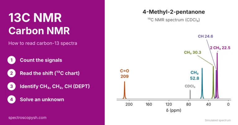
C NMR or H NMR? Use both: proton NMR shows the hydrogens and their neighbours; carbon NMR shows the carbon skeleton.
PART A
How to Read a ¹H NMR Spectrum (Proton NMR)
¹H nuclear magnetic resonance spectroscopy (proton NMR, or HNMR) studies the hydrogen atoms of a molecule. To read an H NMR spectrum, go through four steps in order:
- Count the signals → how many kinds of H
- Read the chemical shift → which kind of H
- Read the integration → how many H of each kind
- Read the splitting → how many neighbouring H
Step 1: Count the Signals (Equivalent Hydrogens)
The number of signals in a ¹H NMR spectrum equals the number of kinds of hydrogen in the molecule. Equivalent hydrogens (same chemical environment) give one signal; hydrogens in different environments give different NMR signals.
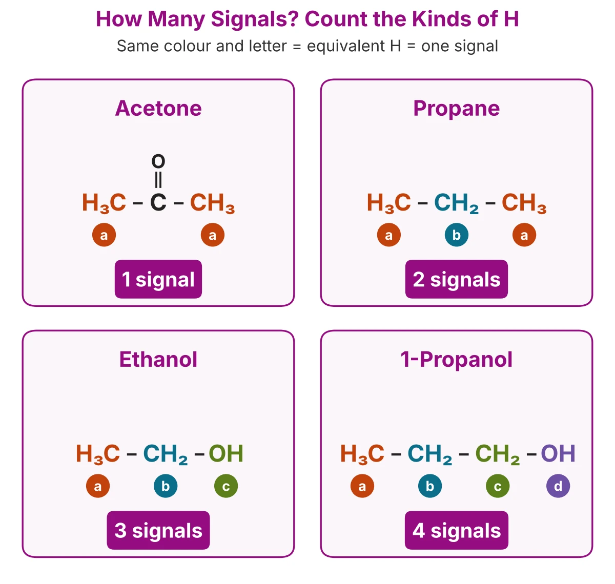
How to Determine the Number of Signals in H NMR
- Hydrogens on the same CH₃ group are equivalent; so are the two H of a simple CH₂ in a chain.
- Look for symmetry. Groups on the two sides of a symmetry plane are equivalent: the two CH₃ groups of acetone give one signal.
- Count every set once, including O–H and N–H hydrogens.
Quick test: replace one H with Cl in your head. If two hydrogens give the same compound when replaced, they are equivalent.
Examples: How Many H NMR Signals?
Step 2: Read the Chemical Shift (Chemical Shift in ¹H NMR)
Chemical shift (δ) is the position of a signal in a nuclear magnetic resonance (NMR) spectrum, measured in parts per million (ppm) from the reference compound tetramethylsilane (TMS) at 0 ppm. It tells you the chemical environment of a hydrogen: which atoms and groups are close to it.
Structure of TMS (Tetramethylsilane)
TMS is the internal reference used to record a proton (¹H) NMR spectrum. A small amount is added to the sample, and its signal is set at δ 0.00 ppm.
- All 12 hydrogens are equivalent, so TMS gives one sharp singlet.
- Its signal appears upfield of almost all organic protons, so it does not overlap with them.
- It is chemically inert and does not react with the sample.
What Do You Understand by the Terms Shielded, Deshielded, Downfield and Upfield in an NMR Spectrum?
Before you study chemical shift values in detail, it helps to understand four terms that describe where a signal appears: shielded, deshielded, downfield and upfield. The ¹H NMR spectrum of acetaldehyde (CH₃CHO) shows all four, because its two signals sit at opposite ends of the spectrum.
Key terms: deshielded vs shielded, downfield vs upfield, explained with the two signals of acetaldehyde:
In short: less electron density → deshielded → downfield; more electron density → shielded → upfield.
Why Chemical Shift Matters
- It identifies the kind of hydrogen: alkyl, next to oxygen, on a double bond, aromatic, aldehyde or acid.
- It is the first clue to the functional groups in an unknown compound.
Each functional group has its own characteristic region in the ¹H NMR spectrum (H NMR regions):
1CH₃ (Methyl Group, sp³ Carbon): 0.85 ppm
A CH₃ on a plain carbon chain is the most shielded common proton.
2CH₃–C=O (Aldehydes, Ketones and Esters): ≈ 2.1 ppm
A CH₃ attached to a carbonyl carbon gives a sharp signal near 2.1 ppm, typical of aldehyde, ketone and ester spectra.
3H–C–O and H–C–X (X = Halogen): 2.5–4.5 ppm
A hydrogen on a carbon that is attached to O or a halogen is deshielded: H–C–Br/I 2.5–4, H–C–Cl 3–4, H–C–O (R–O–CH₃) ≈ 3.3, H–C–F 4–4.5 ppm.
4C=C–H (Vinylic Protons, Alkenes): 4.7–5.3 ppm
Hydrogens on a terminal C=C carbon (R₂C=CH₂) appear near 4.7 ppm; a hydrogen on a more substituted C=C carbon (R₂C=CR–H) appears near 5.3 ppm. A C=C–H next to oxygen (vinyl ethers, =CH–O) is further downfield, near 6.5 ppm.
5Ar–H (Aromatic, Benzene Ring Protons): 6.5–8 ppm
Hydrogens on a benzene ring (aromatic ring protons).
6O=C–H (Aldehyde Proton): 9–10 ppm
A hydrogen on a carbonyl carbon. A signal here strongly suggests an aldehyde.
7COOH (Carboxylic Acid Proton): 10–12 ppm
The most downfield common signal, usually broad.
8O–H and N–H (Alcohols, Phenols, Amines, Amides): variable
Hydrogens on O or N move with solvent and concentration and are often broad singlets.
Solvent and Reference Peaks (CDCl₃ NMR Peak, Water and Impurities)
An NMR sample is dissolved in a deuterated solvent (H replaced by D), because deuterium does not appear in a ¹H NMR spectrum. The solvent is never 100% deuterated, so a small residual solvent peak always appears. Do not count it as a signal of your compound.
- CDCl₃ NMR peak (deuterated chloroform, the most common NMR solvent): residual CHCl₃ at δ 7.26 in ¹H NMR and a triplet at δ 77.16 in ¹³C NMR.
- TMS: δ 0.00 (the reference).
- Water: its position depends on the solvent: ≈ 1.56 in CDCl₃.
Common NMR Solvents: Residual Peaks
Common HNMR Impurities in CDCl₃
Leftover lab solvents give small extra NMR impurity peaks. Check these before you assign a signal:
Tip: a small peak at 7.26 or 1.56 in CDCl₃ is almost always the solvent or water, not your compound.
Step 3: Read the Integration (NMR Integration)
NMR integration is the area under each signal. The area is proportional to the number of hydrogens that give the signal, so integration tells you the ratio of hydrogens in each environment. The NMR instrument shows it as a step curve over each peak or as numbers under the peaks.
What are the red numbers on an H-NMR spectrum? They are usually the integration values: the relative area of each signal, printed under (or above) the peak.
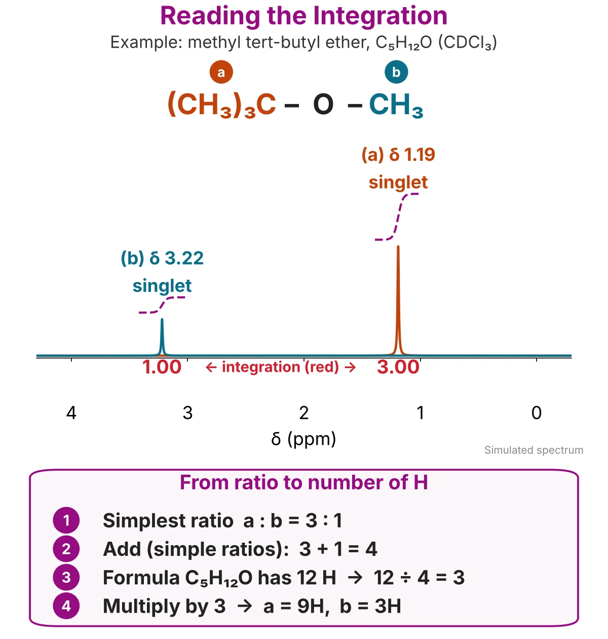
How to Turn the Integration Ratio into Numbers of Hydrogens
- Read the integration value of each signal.
- Divide every value by the smallest one to get the simplest ratio.
- Add the numbers of the ratio and compare the total with the H in the molecular formula.
- Multiply the ratio by (H in formula ÷ total) to get the number of H in each signal.
Integration Measures Area, Not Height
- A broad peak (for example an O–H) can be short but still have a large area. Always read the integration, not the peak height.
- Integration also gives the ratio of two compounds in a mixture: compare the integration per H of one signal from each compound.
Step 4: Read the Splitting (n + 1 Rule)
Splitting (spin–spin coupling) is the division of one signal into several peaks by the hydrogens on the neighbouring carbon. The n + 1 rule: a proton with n equivalent neighbouring hydrogens gives a signal of n + 1 peaks. A signal split into several peaks is called a multiplet.
n = number of neighbouring protons → n + 1 peaks
Singlet, Doublet, Triplet, Quartet and Pascal’s Triangle
The heights of the peaks in a multiplet follow Pascal’s triangle.
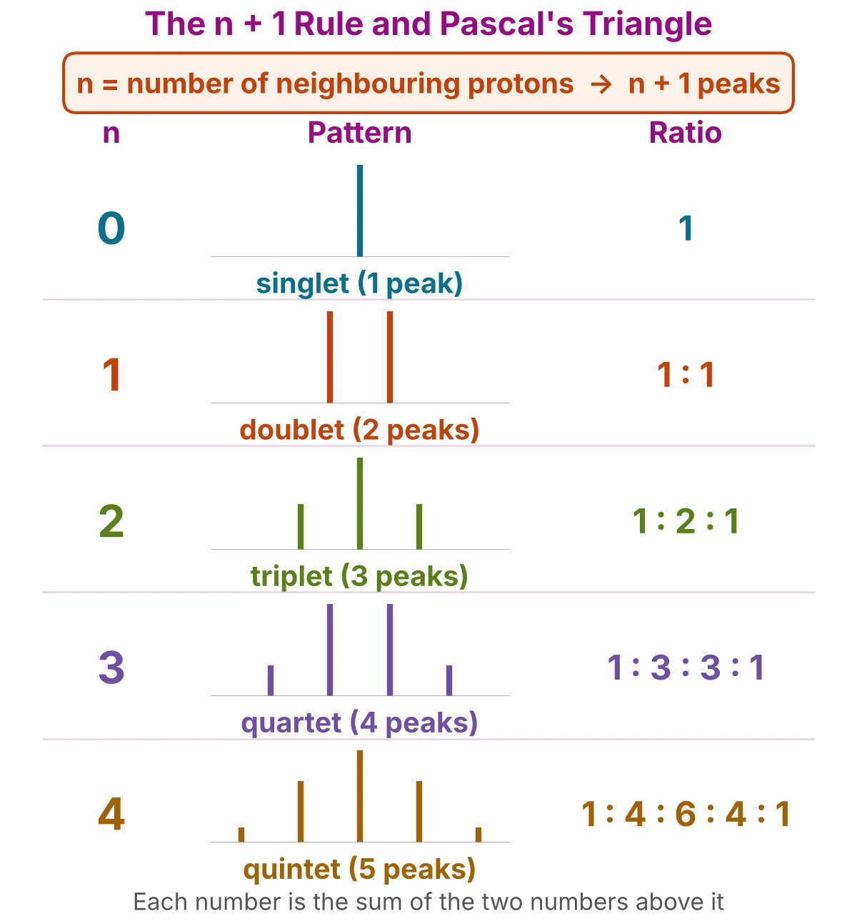
Patterns to remember: a quartet (2H) + triplet (3H) means an ethyl group, –CH₂CH₃; a septet (1H) + doublet (6H) means an isopropyl group, –CH(CH₃)₂.
How to Find the Coupling Constant (J)
The coupling constant (J) is the distance between two neighbouring lines of a multiplet, measured in hertz (Hz). Coupled hydrogens split each other by the same J, so equal J values show which signals are neighbours.
J (Hz) = gap between two lines (ppm) × spectrometer frequency (MHz)
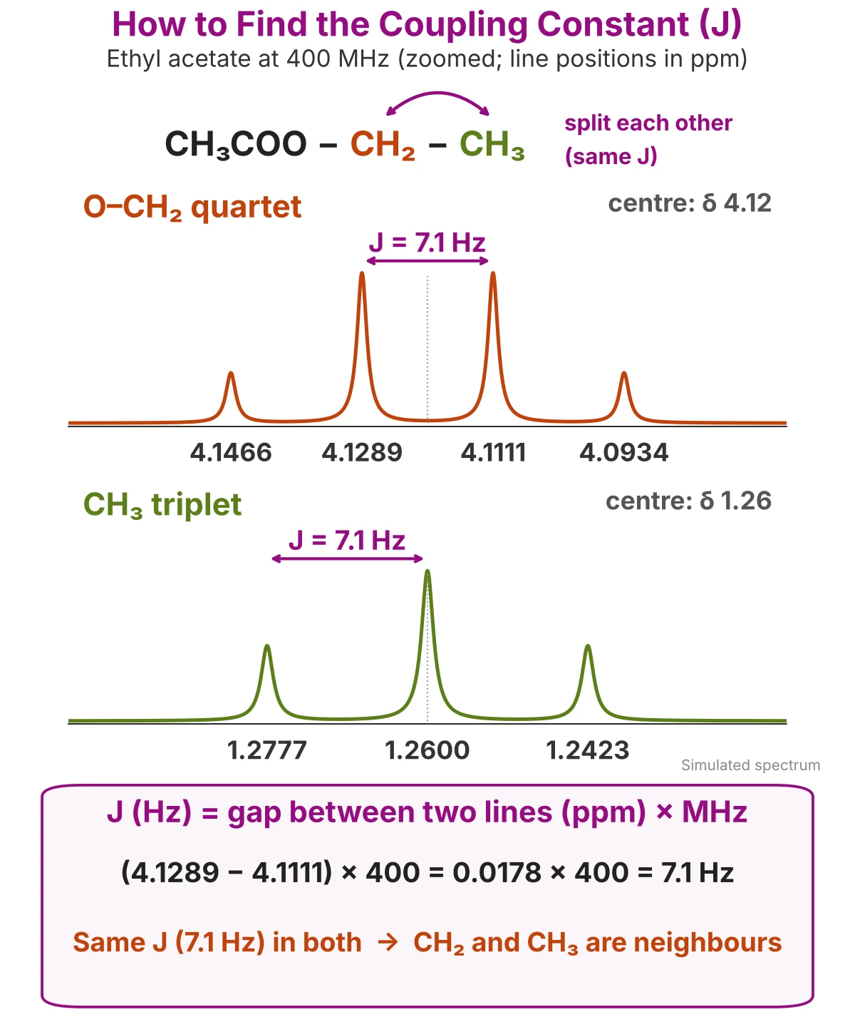
How to calculate the J value in NMR
- Read the δ values (ppm) of two neighbouring lines in the multiplet.
- Subtract them to get the gap in ppm.
- Multiply by the spectrometer frequency in MHz (for example 400).
- The chemical shift of a multiplet is read at its centre (ethyl acetate quartet: δ 4.12).
- Hydrogens usually couple through two or three bonds (H–C–H or H–C–C–H).
Typical Coupling Constants
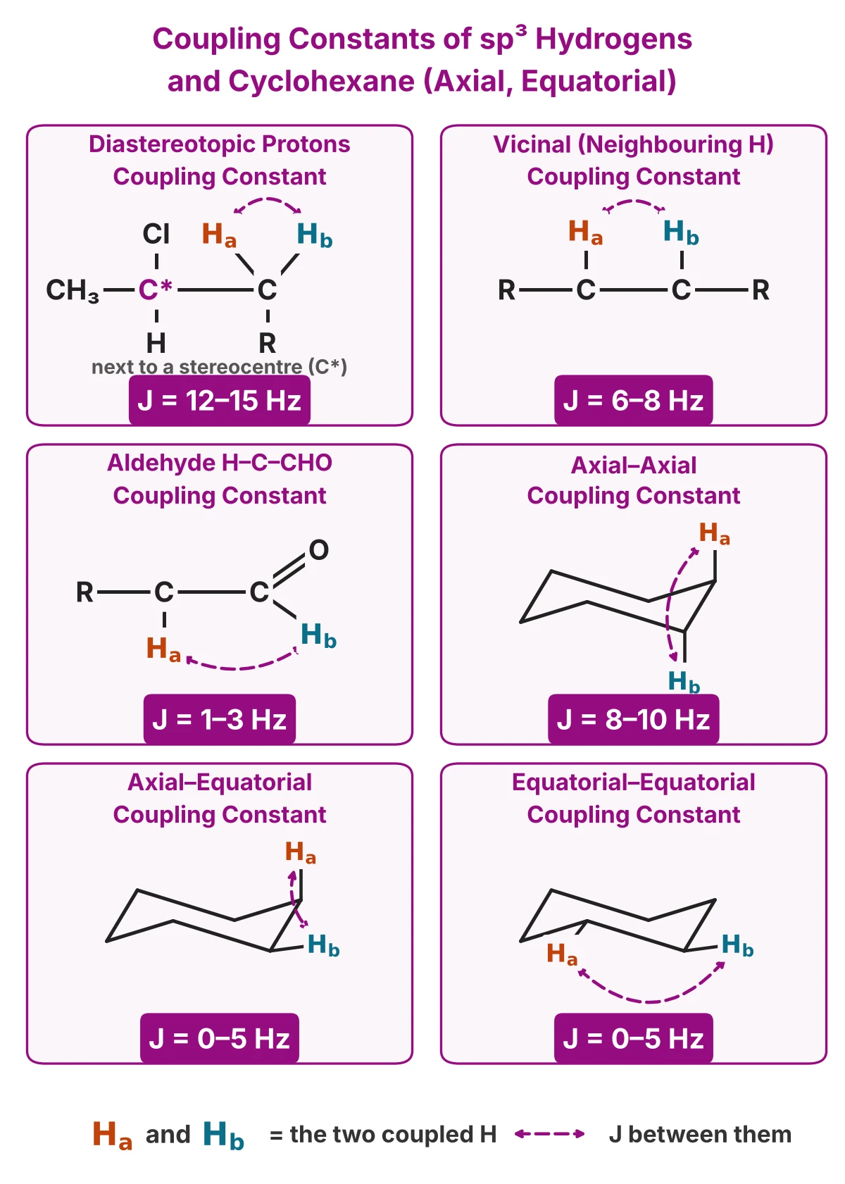
Coupling Constant of Diastereotopic Protons
Two H on the same sp³ carbon (geminal H) split each other only when they are diastereotopic, that is, when they have different chemical shifts (for example a CH₂ next to a stereocentre): J ≈ 12–15 Hz. Equivalent geminal H, such as the two H of a simple CH₂ in a chain, show no splitting from each other.
Vicinal Coupling Constant (Neighbouring Protons)
- H on neighbouring sp³ carbons (H–C–C–H, free rotation): J ≈ 6–8 Hz; for example 7.1 Hz in ethyl acetate.
- Aldehyde H and the neighbouring sp³ H (H–C–CHO): J ≈ 1–3 Hz.
Cyclohexane Coupling Constants: Axial–Axial, Axial–Equatorial and Equatorial–Equatorial
- Axial–axial (trans-diaxial, H–C–C–H at 180°): J ≈ 8–10 Hz, the largest.
- Axial–equatorial and equatorial–equatorial (H–C–C–H at about 60°): J ≈ 0–5 Hz.
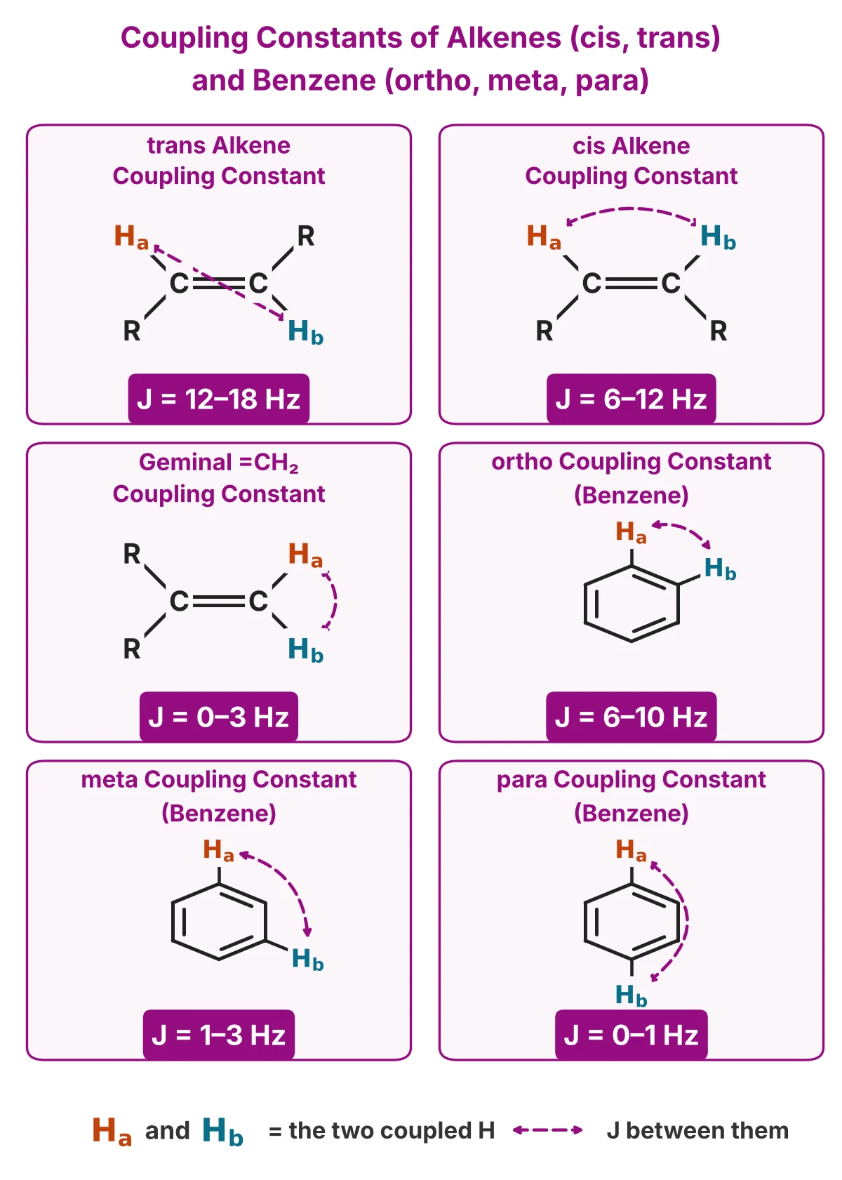
Alkene Coupling Constants: cis, trans and Geminal
- trans H–C=C–H: J ≈ 12–18 Hz
- cis H–C=C–H: J ≈ 6–12 Hz. So J tells a cis alkene from a trans alkene.
- Geminal =CH₂: J ≈ 0–3 Hz
Aromatic Coupling Constants: ortho, meta and para
- ortho (neighbouring ring H): J ≈ 6–10 Hz
- meta: J ≈ 1–3 Hz
- para: J ≈ 0–1 Hz
Coupling Constants Table (Summary)
Doublet of Doublets (dd)
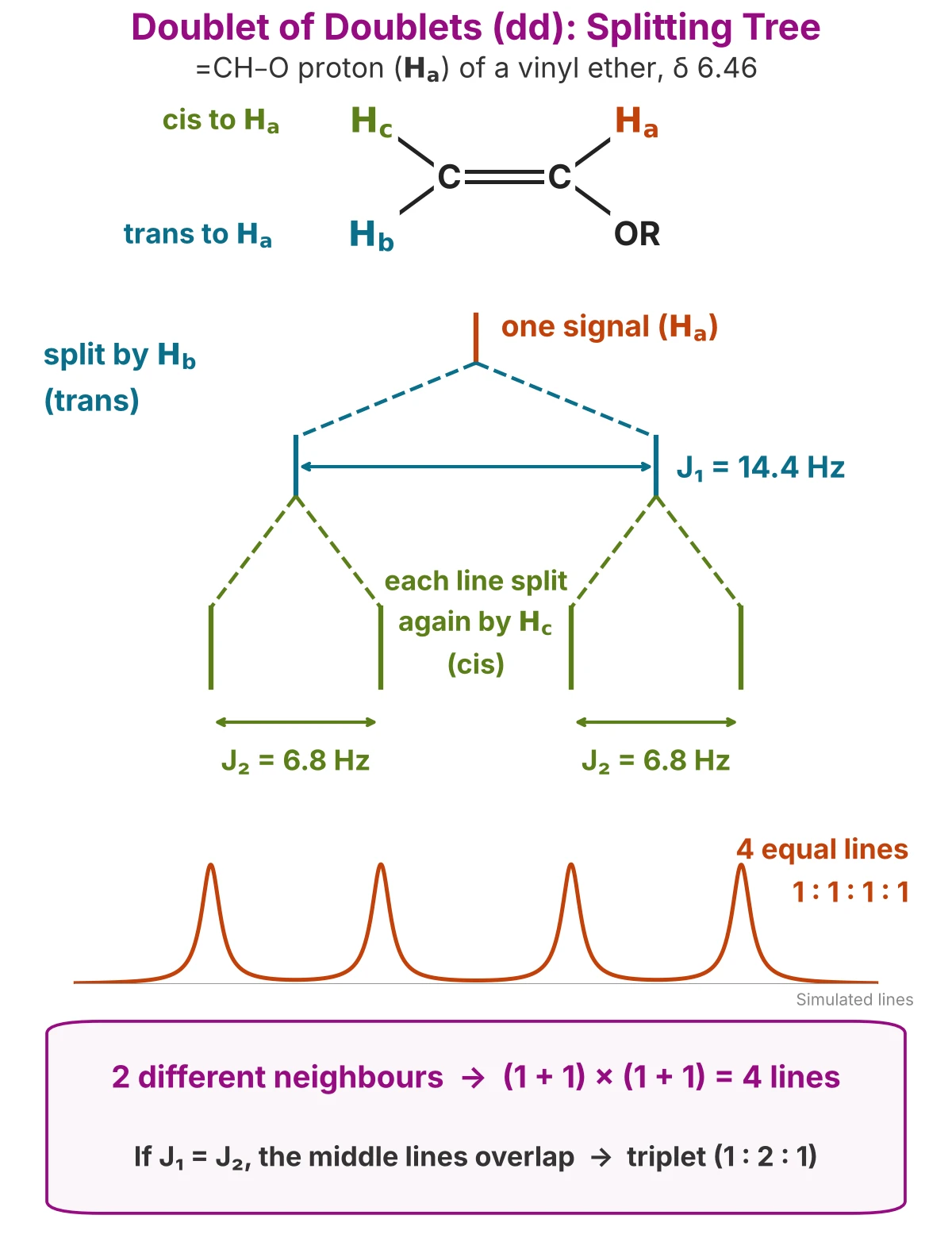
A doublet of doublets (dd) is a signal of four equal lines. It forms when one proton is split by two non-equivalent neighbouring protons with different coupling constants (J₁ ≠ J₂): the first neighbour splits the signal into a doublet, and the second neighbour splits each line again.
2 different neighbours → (1 + 1) × (1 + 1) = 4 lines
How to read a doublet of doublets
- Count four lines of equal height (1 : 1 : 1 : 1).
- The small J (J₂) is the gap between lines 1 and 2 (or 3 and 4).
- The large J (J₁) is the gap between lines 1 and 3 (or 2 and 4).
- The chemical shift is read at the centre of the four lines.
Example: in a vinyl ether (CH₂=CH–O–R), the =CH–O proton at δ 6.46 is a dd with J = 14.4 Hz (trans neighbour) and J = 6.8 Hz (cis neighbour).
Note: if the two J values are equal (J₁ = J₂), the two middle lines overlap and the signal looks like a triplet (1 : 2 : 1), as the n + 1 rule predicts.
Special Cases: Equivalent Hydrogens, OH Peaks and Benzene Rings
The four steps work for most signals. Four special cases explain the signals that do not seem to follow the rules.
1. Equivalent Protons Do Not Split Each Other
Splitting happens only between non-equivalent hydrogens. Equivalent hydrogens on neighbouring carbons give one singlet.
Example: in 1,2-dichloroethane (Cl–CH₂–CH₂–Cl), all 4 H are equivalent, so the spectrum shows one singlet at δ 3.73, not two triplets.
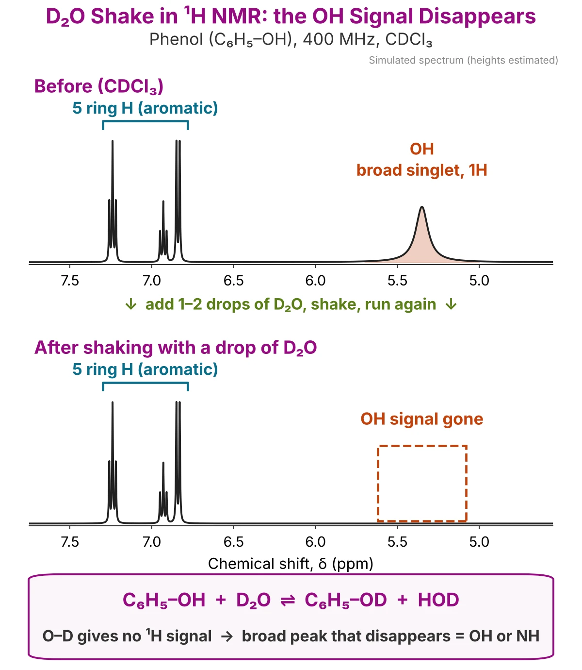
2. OH and NH Protons: Broad Singlet and the D₂O Shake
- Broad singlet: OH and NH hydrogens move quickly between molecules (exchange), so they usually give a broad singlet.
- No splitting: because of this exchange, an OH usually does not split its neighbours, and is not split by them. In ethanol (CDCl₃), the CH₂ at δ 3.72 is a quartet (split by CH₃ only), not split by the OH.
- Position varies: the OH or NH shift changes with concentration, solvent and temperature (alcohol OH about 1–5 ppm, carboxylic acid OH 10–12 ppm).
D₂O shake: broad peak disappears → OH or NH
To confirm an OH or NH signal, add 1–2 drops of D₂O to the NMR tube, shake and run the spectrum again. The OH hydrogen is replaced by deuterium (O–H → O–D), and O–D gives no signal in ¹H NMR, so the peak disappears.
3. Aromatic Protons (Benzene Ring, δ 6.5–8)
- Hydrogens on a benzene ring appear at δ 6.5–8, downfield of most other C–H.
- Ring hydrogens split each other (ortho, meta and para coupling), so they often look like a multiplet (m) rather than a clear doublet or triplet.
- Use the integration of the aromatic region: 5H = monosubstituted ring (phenol, toluene); 4H = disubstituted ring.
4. When the n + 1 Rule Does Not Work (Multiplets)
The n + 1 rule does not give a simple pattern when:
- the neighbours are not equivalent and have different J values (see doublet of doublets);
- the coupled signals are close together in δ: peaks lean and overlap, and the signal is reported as a multiplet (m);
- the hydrogen is an OH or NH that exchanges (broad singlet, see case 2).
¹H NMR Cheat Sheet: How to Read a Proton NMR Spectrum
| Step | Look at | It tells you |
|---|---|---|
| 1 | Number of signals | How many kinds of hydrogen (equivalent H give one signal) |
| 2 | Chemical shift (δ) | The environment: 0.9–1.5 alkyl, ≈2.1 CH₃–C=O, 3.3–4.2 H–C–O, 6.5–8 aromatic, 9–10 CHO, 10–12 COOH |
| 3 | Integration | The relative number of H in each signal (scale the ratio to the molecular formula) |
| 4 | Splitting (n + 1) | Number of neighbouring H: s = 0, d = 1, t = 2, q = 3 |
| J | Coupling constant | Which signals are neighbours (same J); trans 12–18 Hz, cis 6–12 Hz |
- Ethyl group: triplet (3H) + quartet (2H); isopropyl group: doublet (6H) + septet (1H).
- Broad singlet that disappears with D₂O → OH or NH.
- Equivalent H do not split each other; peak area (not height) gives the number of H.
Next: How to Read a ¹³C NMR Spectrum (¹³C chemical shift chart and DEPT). Coming soon.
Worked Example: How to Read the ¹H NMR Spectrum of 2-Hexanone
Let us use the four steps to find the structure of an unknown compound with the molecular formula C₆H₁₂O. Its DBE is 1, so it has one C=O, one C=C or one ring (see the Before You Start box at the top).
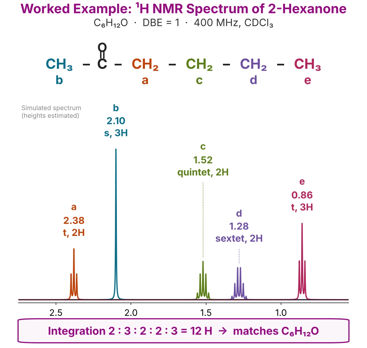
- Step 1: Count the signals. There are 5 signals, so 2-hexanone has 5 kinds of hydrogen (a–e).
- Step 2: Read the chemical shift. The singlet at 2.10 is a CH₃ next to C=O (CH₃–C=O). The triplet at 2.38 is a CH₂ next to C=O. The signals at 0.86–1.52 are alkyl hydrogens. There is no signal at 3.3–4.2 (no H–C–O, so not an alcohol or ether), none at 4.5–6.5 (no C=C–H) and none at 9–10 (no CHO, so not an aldehyde). The one DBE is a ketone C=O.
- Step 3: Read the integration. 2 : 3 : 2 : 2 : 3 = 12 H, which matches C₆H₁₂O. So the groups are two CH₃ and three CH₂.
- Step 4: Read the splitting (n + 1).
- b 2.10 singlet: 0 neighbours → CH₃ on the C=O (methyl ketone).
- a 2.38 triplet: 2 neighbours → CH₂ next to C=O, with one CH₂ on its other side.
- c 1.52 quintet: 4 neighbours → CH₂ between two CH₂.
- d 1.28 sextet: 5 neighbours → CH₂ between a CH₂ and a CH₃.
- e 0.86 triplet: 2 neighbours → CH₃ at the end of the chain.
- All four multiplets share J = 7.5 Hz, so they are neighbours in one chain.
CH₃–CO–CH₂–CH₂–CH₂–CH₃ = 2-hexanone
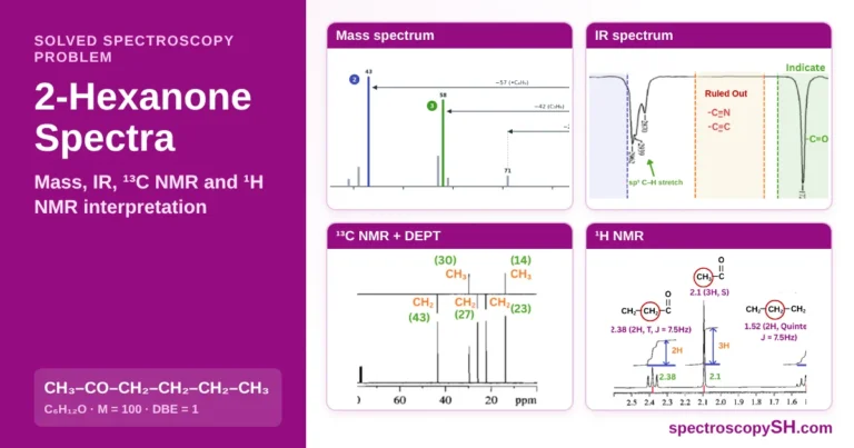
The ¹H NMR spectrum of 2-hexanone shows how the four steps work together: the number of signals, the chemical shift, the integration and the n + 1 splitting each add one piece, and together they give the full structure. For the mass, IR and ¹³C NMR spectra of the same compound, see 2-hexanone spectra interpretation.
Common Mistakes When Reading NMR Spectra
Most errors in reading a ¹H NMR spectrum come from six simple mistakes. Check each one before you draw the final structure.
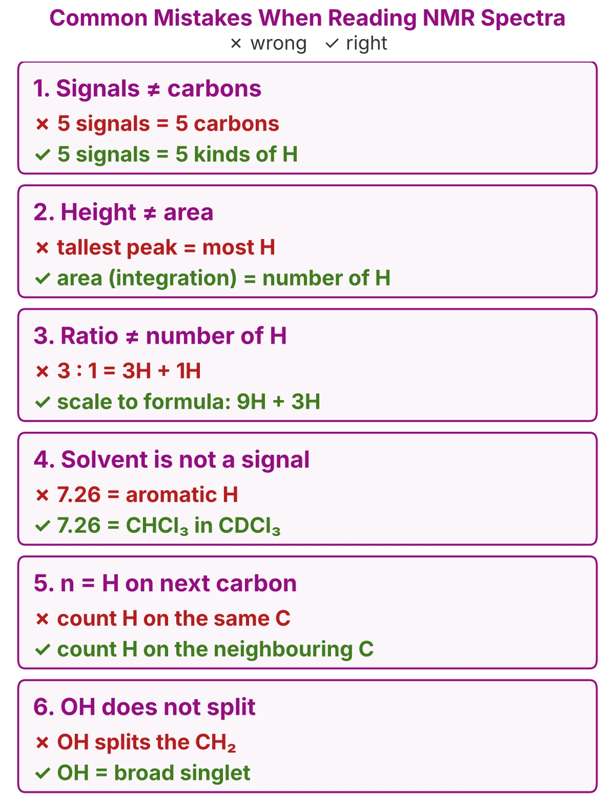
- 1. Counting signals as carbons. A ¹H NMR signal is a kind of hydrogen, not a carbon. 2-hexanone has 6 carbons but only 5 signals, because the C=O carbon has no hydrogen. Step 1
- 2. Reading peak height instead of area. The number of hydrogens is given by the area (integration), not the height. A sextet is short because its area is shared among six lines, but it still counts 2H. Step 3
- 3. Taking the integration ratio as the number of H. Integration gives a ratio. Scale it to the molecular formula: a 3 : 1 ratio in C₅H₁₂O (methyl tert-butyl ether) means 9H : 3H. Step 3
- 4. Counting the solvent peak as a signal. In CDCl₃, the peak at 7.26 is the small amount of CHCl₃ in the solvent, and the peak near 1.56 is water. They are not part of your compound. Solvent peaks
- 5. Counting the wrong hydrogens in n + 1. n is the number of H on the neighbouring carbon(s), not the H on the same carbon. In ethyl acetate, the CH₃ at 2.05 is a singlet: the carbon next to it is the C=O, which has no hydrogen. Step 4
- 6. Expecting an OH to split its neighbours. An OH usually gives a broad singlet and does not split the CH next to it; in ethanol (CDCl₃) the CH₂ is a quartet, split by the CH₃ only. Confirm an OH with the D₂O shake. Special cases
How to Read NMR Spectra: Practice MCQs with Answers
Fifteen questions on reading a ¹H NMR spectrum: the first ten test each step, the last five test the ideas behind them. Pick an answer, then open it to check.
1. Proton NMR (¹H NMR), how to determine the number of signals: how many signals does ethanol (CH₃CH₂OH) give in its ¹H NMR spectrum?
- 2
- 3
- 4
- 5
- 6
Show answer
Answer: B. Ethanol has three kinds of hydrogen: CH₃, CH₂ and OH, so its ¹H NMR spectrum shows 3 signals. (Step 1)
2. Downfield vs upfield on an H NMR chart: which statement is correct?
- Downfield is on the right, near TMS
- Downfield signals have larger δ and appear on the left
- Upfield signals are more deshielded
- TMS appears at 7.26 ppm
- δ increases from left to right
Show answer
Answer: B. δ increases from right to left. Signals on the left (larger δ) are downfield and deshielded; TMS is at 0.00 ppm on the far right. (What Is an NMR Spectrum?)
3. Using an H NMR shift table, a singlet at δ 9.8 ppm in a ¹H NMR spectrum most likely belongs to:
- A CH₃ next to C=O
- An aromatic H
- A carboxylic acid OH
- An alkyl CH₃
- An aldehyde H (CHO)
Show answer
Answer: E. Aldehyde hydrogens appear at 9–10 ppm; acetaldehyde’s CHO is at 9.79 ppm. (Step 2)
4. Integration in nuclear magnetic resonance (NMR) spectroscopy: methyl tert-butyl ether (C₅H₁₂O) shows two singlets in the ratio 3 : 1. How many hydrogens give each signal?
- 9H and 3H
- 3H and 1H
- 6H and 2H
- 12H and 4H
- 3H and 3H
Show answer
Answer: A. Integration gives a ratio. The formula has 12 H, so 3 : 1 is scaled ×3 to 9H (three CH₃) and 3H (OCH₃). (Step 3)
5. By the n + 1 rule in NMR, the CH₂ signal of an ethyl group (–CH₂–CH₃) appears as a:
- Singlet
- Doublet
- Triplet
- Quartet
- Quintet
Show answer
Answer: D. The CH₂ has 3 neighbouring hydrogens (the CH₃), so n + 1 = 4 lines: a quartet. (Step 4)
6. How to find the coupling constant from nuclear magnetic resonance spectra: two neighbouring lines of a multiplet are at 4.1289 and 4.1111 ppm on a 400 MHz spectrometer. What is J?
- 0.018 Hz
- 1.8 Hz
- 7.1 Hz
- 17.8 Hz
- 71 Hz
Show answer
Answer: C. J = Δδ (ppm) × spectrometer frequency (MHz) = 0.0178 × 400 = 7.1 Hz. (Coupling constant J)
7. Typical coupling constants: a J of 15 Hz between two hydrogens on a C=C double bond shows that they are:
- cis
- trans
- Geminal (=CH₂)
- ortho on a benzene ring
- para on a benzene ring
Show answer
Answer: B. Alkene trans coupling is 12–18 Hz; cis is 6–12 Hz and geminal =CH₂ is 0–3 Hz. (Typical coupling constants)
8. Doublet of doublets NMR: one proton is split by two different neighbouring protons with different coupling constants (J₁ ≠ J₂). How many lines does its signal show?
- 2
- 3
- 4
- 5
- 6
Show answer
Answer: C. Each neighbour doubles the lines: (1 + 1) × (1 + 1) = 4 equal lines, a doublet of doublets (dd). (Doublet of doublets)
9. Broad singlet OH in H NMR: in the ¹H NMR spectrum of phenol, a broad singlet at δ 5.35 ppm disappears after shaking the sample with D₂O. This signal is:
- An aromatic H
- The CDCl₃ solvent peak
- The OH proton
- A CH₃ group
- TMS
Show answer
Answer: C. D₂O exchanges the OH hydrogen for deuterium (O–H → O–D), and O–D gives no ¹H signal, so the OH peak disappears. (Special cases)
10. NMR solvent impurities and the CDCl₃ NMR peak: in a ¹H NMR spectrum recorded in CDCl₃, a small singlet at 7.26 ppm comes from:
- Aromatic H of the compound
- Water
- TMS
- Residual CHCl₃ in the solvent
- Grease
Show answer
Answer: D. CDCl₃ always contains a little CHCl₃, which gives a singlet at 7.26 ppm. It is not a signal of your compound. (Solvent peaks)
11. Chemical shift NMR concept: why does the O–CH₂ of ethyl acetate appear at 4.12 ppm, but its CH₃ at 1.26 ppm?
- The CH₂ has fewer hydrogens
- The CH₂ signal is a quartet
- Oxygen pulls electron density away from the CH₂ hydrogens, so they are deshielded and appear downfield
- Oxygen adds electron density to the CH₂, so it is shielded
- The CH₃ is closer to the solvent peak
Show answer
Answer: C. An electronegative atom (O) pulls electron density away from nearby hydrogens. Less shielded (deshielded) hydrogens appear at larger δ, downfield. (Step 2)
12. NMR spectrometer frequency: a signal is at δ 2.0 ppm on a 400 MHz instrument. Where does it appear on a 300 MHz instrument?
- 1.5 ppm
- 2.0 ppm
- 2.7 ppm
- 600 ppm
- 800 ppm
Show answer
Answer: B. The ppm scale does not depend on the instrument: δ stays 2.0 ppm. Only the distance from TMS in Hz changes (800 Hz at 400 MHz, 600 Hz at 300 MHz). (What Is an NMR Spectrum?)
13. Splitting pattern NMR: a ¹H NMR spectrum shows a doublet (6H) and a septet (1H) with the same J. Which group is present?
- Ethyl, –CH₂CH₃
- tert-Butyl, –C(CH₃)₃
- Methoxy, –OCH₃
- n-Propyl, –CH₂CH₂CH₃
- Isopropyl, –CH(CH₃)₂
Show answer
Answer: E. The 6H doublet is two equivalent CH₃ with one neighbour (the CH); the 1H septet is the CH with six neighbours (n + 1 = 7). Same J shows they split each other. (Step 4)
14. Equivalent protons in NMR: why does 1,2-dichloroethane (Cl–CH₂–CH₂–Cl) show only one singlet, although each CH₂ has two hydrogens on the next carbon?
- All four hydrogens are equivalent, and equivalent hydrogens do not split each other
- Chlorine stops splitting
- The signal is too broad to show splitting
- The hydrogens exchange with the solvent
- The two CH₂ groups are too far apart
Show answer
Answer: A. Both CH₂ groups are the same by symmetry, so all 4 H are equivalent. Splitting happens only between non-equivalent hydrogens, so the signal is a singlet (δ 3.73). (Special cases)
15. NMR spectroscopy problem: a compound C₂H₄O₂ shows a singlet at 2.10 ppm (3H) and a broad singlet near 11.5 ppm (1H) that disappears with D₂O. What is it?
- Methyl formate, HCOOCH₃
- Acetaldehyde, CH₃CHO
- Ethanol, CH₃CH₂OH
- Acetic acid, CH₃COOH
- Acetone, CH₃COCH₃
Show answer
Answer: D. The broad 1H signal at 10–12 ppm that is lost with D₂O is a COOH; the 3H singlet near 2.1 is a CH₃ next to C=O. Methyl formate (also C₂H₄O₂) would show a CHO singlet near 8 and an OCH₃ singlet near 3.8. (Steps 1–4)
How to Read NMR Spectra: Frequently Asked Questions
How do you read an NMR spectrum step by step?
Answer four questions in order: how many signals (kinds of hydrogen), where they are (chemical shift, δ), how big they are (integration, number of H) and how they are split (n + 1 rule, number of neighbouring H). Then join the pieces into a structure that fits the molecular formula.
What is NMR?
Nuclear magnetic resonance (NMR) is a method that uses a strong magnet and radio waves to study the nuclei of atoms such as ¹H and ¹³C. An NMR spectrum shows how many kinds of hydrogen or carbon a molecule has and how they are joined.
What is the principle of NMR (how does NMR work)?
Nuclei such as ¹H behave like tiny magnets. In a strong magnetic field, a pulse of radio waves makes them absorb energy (resonance), and the exact frequency they respond to depends on their chemical environment, which the instrument records as the spectrum.
What are the two main types of NMR?
The two main types used in organic chemistry are ¹H NMR (proton NMR), which studies hydrogens, and ¹³C NMR (carbon-13 NMR), which studies carbons. Other nuclei, such as ¹⁹F and ³¹P, can also be studied.
What is NMR used for?
Nuclear magnetic resonance (NMR) spectroscopy is used to find the structure of organic compounds, check their purity, identify impurities and measure the ratio of compounds in a mixture.
What is NMR vs MRI?
Both use the same principle. NMR spectroscopy gives a spectrum that shows the structure of a molecule, while MRI (magnetic resonance imaging) uses the NMR signal of hydrogen, mostly in water and fat, to make pictures of the inside of the body.
What type of radiation is used in NMR?
NMR uses radio waves (radio-frequency electromagnetic radiation), the lowest-energy radiation used in spectroscopy. Their wavelength is about 0.5–1 m on common 300–600 MHz instruments.
Which frequency is used in NMR?
It depends on the strength of the magnet. A “400 MHz” instrument detects ¹H at about 400 MHz and ¹³C at about 100 MHz; common instruments work at 300–600 MHz for ¹H.
What instrument is used for NMR spectroscopy?
An NMR spectrometer: a strong superconducting magnet (cooled with liquid helium), a probe that holds the sample tube, a radio-frequency transmitter and receiver, and a computer that turns the signal into a spectrum.
What is the standard used in NMR spectroscopy?
Tetramethylsilane (TMS), Si(CH₃)₄, is the standard and is set at 0.00 ppm. Its 12 equivalent hydrogens give one sharp singlet upfield of almost all other signals; the residual solvent peak (7.26 ppm in CDCl₃) is also often used as a reference.
What increases ppm in NMR?
Deshielding increases the ppm value (moves a signal downfield). Hydrogens near electronegative atoms (O, N, halogens), next to a C=O, or on a C=C or benzene ring are deshielded and appear at higher ppm.
What are the red numbers on an H NMR spectrum?
They are usually the integration values printed under each signal. They give the relative number of hydrogens in each signal, which you scale to the molecular formula.
What does 5H integration mean in H NMR?
It means the signal comes from five hydrogens. A 5H signal at 6.5–8 ppm usually shows a monosubstituted benzene ring, as in phenol or toluene.
What is the n + 1 rule in NMR?
A hydrogen with n equivalent hydrogens on the neighbouring carbon(s) gives a signal split into n + 1 lines. Example: the CH₃ of an ethyl group has 2 neighbours, so it is a triplet (2 + 1 = 3). See Step 4.
C NMR or H NMR: which one should you use?
Use both: ¹H NMR shows the kinds of hydrogen, how many there are and their neighbours, while ¹³C NMR counts the kinds of carbon, including carbons with no hydrogen such as C=O. Start with ¹H NMR and confirm the carbon skeleton with ¹³C NMR (see Proton NMR vs Carbon NMR).
What solvents are used in NMR?
Deuterated solvents, in which hydrogen is replaced by deuterium so they give no large ¹H signal: CDCl₃ (the most common), DMSO-d₆, acetone-d₆, CD₃OD, C₆D₆ and D₂O. Each still shows a small residual peak (see the solvent peaks table above).
Why is CDCl₃ used in NMR?
CDCl₃ dissolves most organic compounds, and its deuterium gives no signal in ¹H NMR, so the solvent does not hide the sample’s peaks. Only the small amount of CHCl₃ left in it shows a peak, at 7.26 ppm.
Why is D₂O used in NMR?
D₂O is used as the solvent for water-soluble compounds such as sugars, amino acids and salts. A few drops are also added in the D₂O shake test: OH and NH hydrogens exchange for deuterium and their signals disappear.
What is the difference between upfield and downfield in NMR?
Downfield signals are on the left (larger δ): their hydrogens are deshielded, for example near oxygen or a C=O. Upfield signals are on the right (smaller δ, near TMS at 0 ppm): their hydrogens are shielded, as in alkyl groups.
What is a doublet of doublets in NMR?
A doublet of doublets (dd) is a signal of four equal lines from one hydrogen split by two different neighbouring hydrogens with different coupling constants. Example: the =CH–O proton of a vinyl ether at 6.46 ppm, with J = 14.4 and 6.8 Hz (see doublet of doublets).
What are the limitations of NMR?
NMR needs milligram amounts of sample (it is much less sensitive than mass spectrometry), uses an expensive instrument and deuterated solvents, and signals can overlap in large molecules. ¹³C NMR is slow because only 1.1% of carbon is ¹³C.
How to Read NMR Spectra: Key Terms
Chemical shift (δ): the distance of a signal from the TMS signal (0.00 ppm), measured in ppm; it shows the type of hydrogen. Example: the CHO hydrogen of acetaldehyde is at 9.79 ppm.
Tetramethylsilane (TMS): the reference compound, Si(CH₃)₄, whose signal is set at 0.00 ppm. Example: all other ¹H signals are measured from the TMS singlet at 0.00 ppm.
Downfield and upfield: downfield is the left of the spectrum (larger δ); upfield is the right (smaller δ, near TMS). Example: the O–CH₂ of ethyl acetate (4.12 ppm) is downfield of its CH₃ (1.26 ppm).
Shielding and deshielding: electrons around a hydrogen shield it and move its signal upfield; removing electron density deshields it and moves the signal downfield. Example: oxygen deshields the O–CH₂ hydrogens of ethyl acetate.
Equivalent protons: hydrogens in the same chemical environment; they give one signal and do not split each other. Example: the 4 H of Cl–CH₂–CH₂–Cl give one singlet at 3.73 ppm.
Integration: the area under a ¹H NMR signal; it gives the relative number of hydrogens. Example: methyl tert-butyl ether shows 9H and 3H (ratio 3 : 1).
n + 1 rule: n equivalent hydrogens on the neighbouring carbon split a signal into n + 1 lines. Example: the CH₃ of an ethyl group has 2 neighbours, so it is a triplet.
Multiplicity: the number of lines in a signal: singlet (s), doublet (d), triplet (t), quartet (q) or multiplet (m). Example: the CH₃ of acetone is a singlet at 2.17 ppm.
Coupling constant (J): the distance in Hz between two lines of a split signal; signals that split each other have the same J. Example: J = 7.1 Hz for the quartet and triplet of ethyl acetate.
Doublet of doublets (dd): four equal lines from one hydrogen split by two different neighbours with different J. Example: the =CH–O hydrogen of a vinyl ether: J = 14.4 and 6.8 Hz.
Diastereotopic protons: two hydrogens on the same CH₂ that are not equivalent, usually because of a nearby stereocentre; they can split each other. Example: the two H of the CH₂ in 2-butanol, next to its stereocentre.
Deuterated solvent: a solvent in which H is replaced by deuterium (D), so it gives no large ¹H signal. Example: CDCl₃, with a small residual CHCl₃ peak at 7.26 ppm.
D₂O shake: adding D₂O to the sample: OH and NH hydrogens exchange for D and their signals disappear. Example: the broad OH singlet of phenol at 5.35 ppm disappears.
Spectrometer frequency (MHz): the frequency at which the instrument detects ¹H; it sets how many Hz one ppm equals. Example: on a 400 MHz instrument, 1 ppm = 400 Hz.
Learn More: NMR, IR and Mass Spectra Worked Examples
Read: worked examples with ¹H NMR
2-Hexanone Spectra (Mass, IR, ¹³C and ¹H NMR) Ethyl Acetate Spectra and Structure Elucidation
Read: other spectroscopy
IR Spectrum Interpretation Mass Spectrum Interpretation What is Mass Spectrometry? Base Peak and Molecular Ion Peak
Try: free tools
IR Peak Identifier IR Spectrum Comparison Tool Mass Spectrum Fragment Identifier Molecular Mass Calculator
Identify the same compounds by chemical tests on chemistrySH.com
Phenol Identification Tests Identification of Acetone Identification of Acetaldehyde Identification of Benzaldehyde Iodoform Test for Methyl Ketones chemistrySH.com
Sources and Further Reading
[1] Bruice, P. Y. (2016). Organic chemistry (8th ed.). Pearson.
[2] Gottlieb, H. E., Kotlyar, V., & Nudelman, A. (1997). NMR chemical shifts of common laboratory solvents as trace impurities. The Journal of Organic Chemistry, 62(21), 7512–7515. https://doi.org/10.1021/jo971176v
[3] Fulmer, G. R., Miller, A. J. M., Sherden, N. H., Gottlieb, H. E., Nudelman, A., Stoltz, B. M., Bercaw, J. E., & Goldberg, K. I. (2010). NMR chemical shifts of trace impurities: Common laboratory solvents, organics, and gases in deuterated solvents relevant to the organometallic chemist. Organometallics, 29(9), 2176–2179. https://doi.org/10.1021/om100106e
[4] Saikia, E., Bora, S. J., & Chetia, B. (2015). H₂O₂ in WERSA: An efficient green protocol for ipso-hydroxylation of aryl/heteroarylboronic acid. RSC Advances, 5(124), 102723–102726. https://doi.org/10.1039/C5RA21354A