13C NMR (Carbon NMR): How to Read Carbon-13 NMR Spectra Step by Step
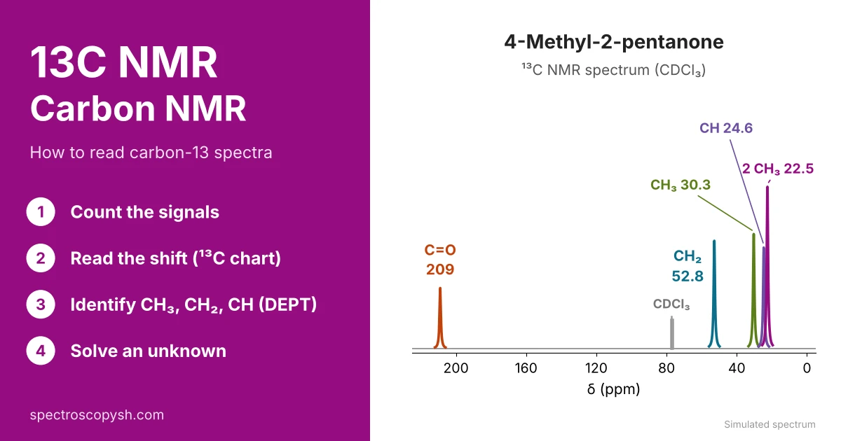
13C NMR (carbon NMR, also written C13 NMR, C-13 NMR or CNMR) is the nuclear magnetic resonance spectrum of the carbon-13 atoms in a molecule, so it shows the carbon skeleton. To read a 13C NMR spectrum, answer three questions: how many signals (kinds of carbon), where they appear (chemical shift, δ) and what type each carbon is (CH₃, CH₂, CH or a carbon with no hydrogen, from DEPT). Each answer gives one piece of the structure.
What Information Can Be Obtained from a ¹³C NMR Spectrum?
1. Number of Signals
Tells you how many kinds of carbon (chemically different carbons) are in the molecule.
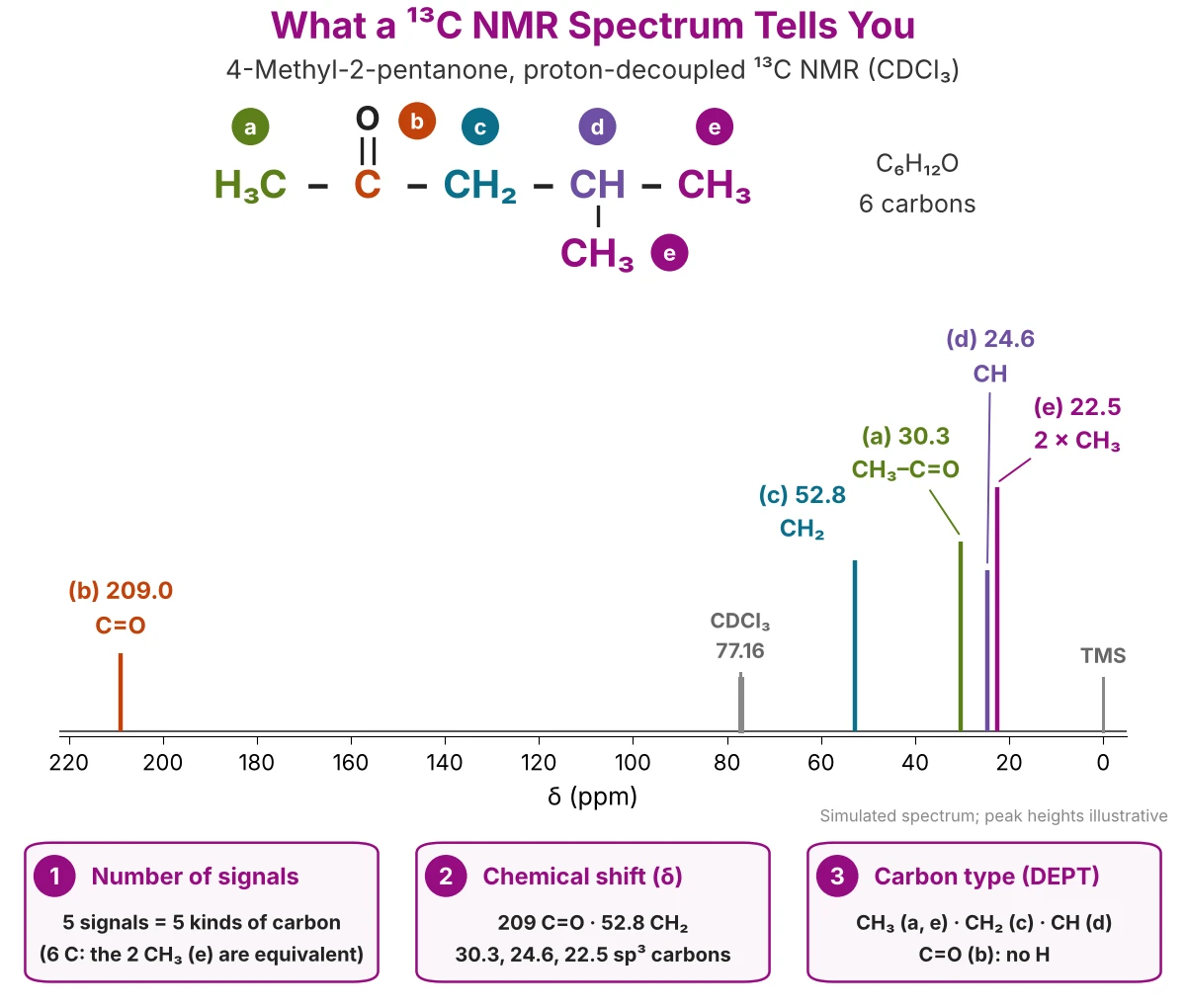
5 signals = 5 kinds of carbon. Equivalent carbons give one signal, so the number of signals can be smaller than the number of carbons.
2. Chemical Shift (δ)
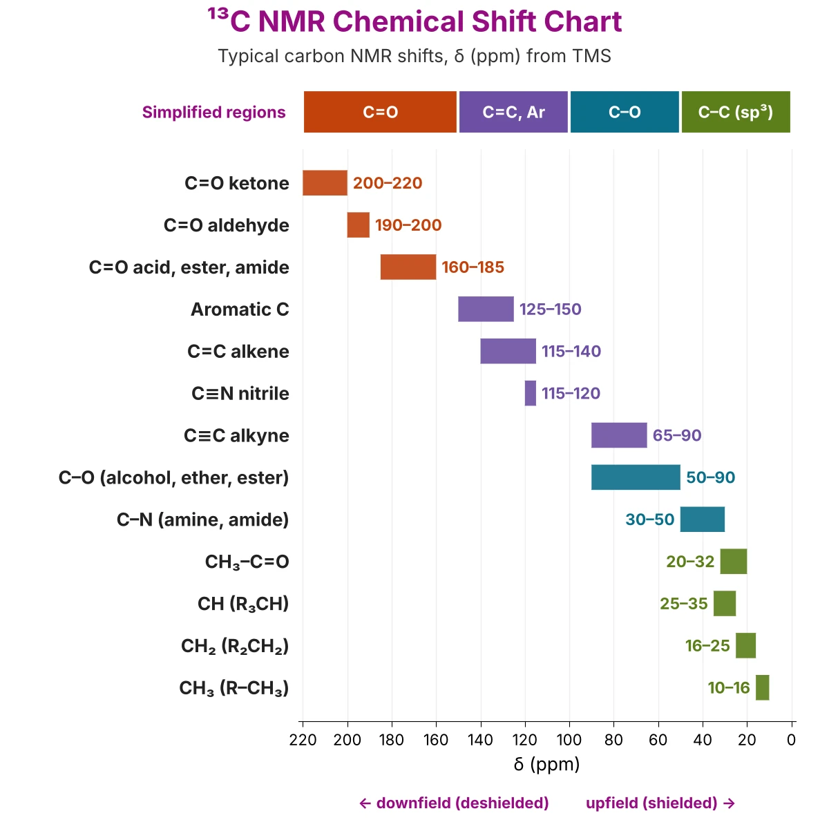
Tells you which type of carbon is present. ¹³C chemical shifts cover about 0–220 ppm, so signals rarely overlap. For example:
- 0–50 ppm → sp³ carbons (CH₃, CH₂, CH)
- 50–90 ppm → carbon bonded to oxygen (C–O)
- 100–150 ppm → alkene (C=C) and aromatic ring carbons
- 160–185 ppm → C=O of carboxylic acids, esters and amides
- 190–220 ppm → C=O of aldehydes and ketones
3. Type of Carbon (DEPT)
Tells you how many hydrogens are on each carbon. The DEPT NMR spectra give this:
- DEPT-135 → CH₃ and CH point up, CH₂ points down
- DEPT-90 → only CH appears
- Carbon with no hydrogen (C=O, quaternary C) → absent in DEPT; seen only in the normal ¹³C spectrum
What a routine ¹³C NMR spectrum does not show: the usual spectrum is proton-decoupled, so every kind of carbon gives one sharp line (no splitting), and peak heights are not used to count carbons (no integration). See proton-coupled and proton-decoupled ¹³C NMR spectra.
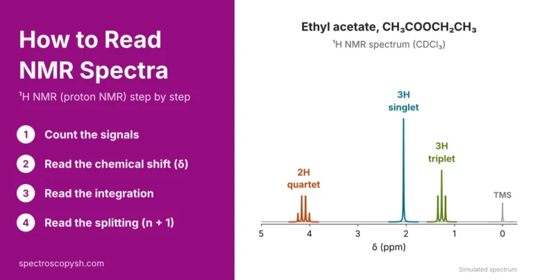
¹³C NMR shows the carbons; ¹H NMR shows the hydrogens on them, with integration and splitting (n + 1 rule). For the hydrogen side, see How to Read NMR Spectra: ¹H NMR step by step.
What Is a ¹³C NMR Spectrum?
A ¹³C NMR spectrum (C NMR spectrum or CNMR spectrum) is a graph of the signals given by the carbon-13 atoms of a molecule. The routine spectrum is proton-decoupled, so each kind of carbon gives one sharp line, called a peak. Figure 1 shows a typical example.
How to Read the Axes and Peaks
- Horizontal axis: chemical shift, δ (ppm), from about 0 to 220 ppm, increasing from right to left. Left = downfield (deshielded carbon); right = upfield (shielded carbon).
- Reference: TMS (tetramethylsilane) is set at 0.00 ppm.
- Solvent: CDCl₃ gives a small triplet at 77.16 ppm; it is not part of the sample (see solvent peaks in ¹³C NMR).
- Peaks: one line for each kind of carbon. Peak height is not proportional to the number of carbons, so ¹³C NMR peaks are counted, not measured.
Why Carbon-13? The C-13 Isotope
Carbon has two stable isotopes. Carbon-12 (98.9 %) has a nuclear spin of zero, so it gives no NMR signal. Carbon-13, the C-13 isotope, makes up only about 1.1 % of natural carbon; its nucleus has 6 protons and 7 neutrons and a spin of ½, like ¹H, so it is NMR active. ¹³C NMR therefore sees only about 1 carbon atom in 90.
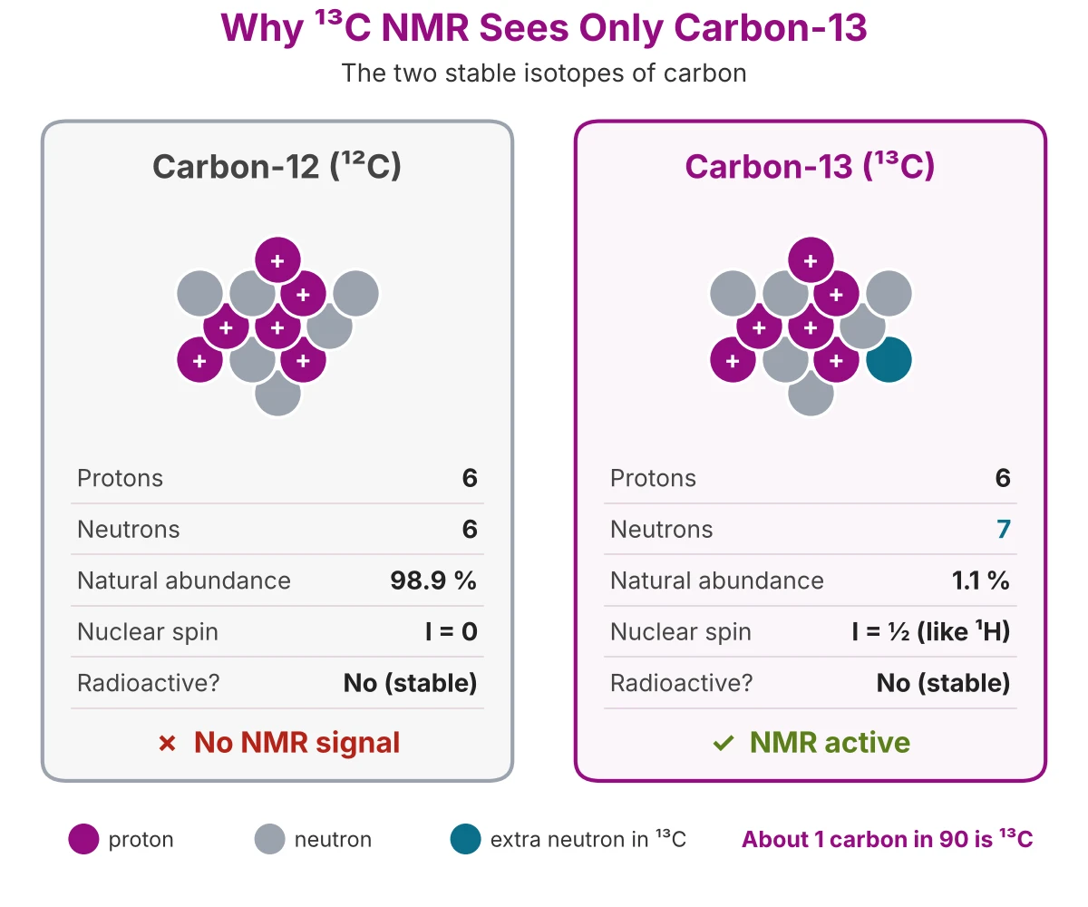
Carbon-12, Carbon-13 and Carbon-14 Compared
All three have 6 protons; only ¹³C has a nuclear spin (½), so only ¹³C gives an NMR signal. *Carbon-14 is radioactive (half-life ≈ 5,730 years); carbon-12 and carbon-13 are stable.
Why ¹³C NMR Signals Are Weak
- Low abundance: only 1.1 % of the carbon atoms are ¹³C.
- Weak magnet: the ¹³C nucleus resonates at about one-fourth the frequency of ¹H (about 100 MHz on a 400 MHz instrument), and its signal is about 6000 times weaker than that of ¹H.
- Result: a ¹³C spectrum needs more sample and many scans added together, so it takes longer to record than a ¹H spectrum.
Useful side effect: because ¹³C atoms are rare, two ¹³C atoms are almost never next to each other in one molecule, so ¹³C signals are not split by neighbouring carbons. The same 1.1 % of ¹³C also causes the small M + 1 peak in a mass spectrum.
¹³C NMR vs ¹H NMR (C NMR or H NMR?)
C NMR or H NMR? Use both. ¹H NMR (proton NMR) shows the hydrogens, how many there are and their neighbours; ¹³C NMR (carbon NMR) shows the carbon skeleton, including carbons that carry no hydrogen. The main differences between HNMR and CNMR are:
Same Molecule, Two Spectra: 4-Methyl-2-pentanone
The table puts the ¹H and ¹³C NMR data of 4-methyl-2-pentanone (Figure 1) side by side. The letters a–e match the structure in Figure 1.
What each spectrum adds: ¹³C NMR shows the C=O carbon (209.0 ppm), which has no hydrogen and so gives no ¹H signal, and it separates the CH₃–C=O and CH carbons that overlap at 2.12–2.13 ppm in ¹H NMR. ¹H NMR adds what ¹³C NMR cannot: the number of hydrogens (integration) and the neighbours (splitting). To read the hydrogen side, see How to Read NMR Spectra (¹H NMR).
Step 1: Count the Signals (How Many ¹³C NMR Signals?)
In a proton-decoupled ¹³C NMR spectrum, each kind of carbon gives one signal (one line). Count the lines, but leave out TMS (0 ppm) and the solvent triplet (CDCl₃, 77.16 ppm).
Number of ¹³C signals = number of kinds of carbon
Equivalent Carbons
Carbons are equivalent when the symmetry of the molecule (a mirror plane or a rotation) exchanges them. Equivalent carbons are in the same environment, so they give one signal. Quick rules:
- Groups on the same carbon: the two CH₃ of an isopropyl group, (CH₃)₂CH–, give one signal; the three CH₃ of a tert-butyl group, (CH₃)₃C–, give one signal.
- Mirror plane: in 3-pentanone, CH₃CH₂COCH₂CH₃, the two ethyl groups match, so 5 carbons give 3 signals.
- Benzene rings: benzene gives 1 signal; a monosubstituted ring (C₆H₅–X) gives 4 ring signals; a para-disubstituted ring with two identical groups gives 2 ring signals (with two different groups, 4).
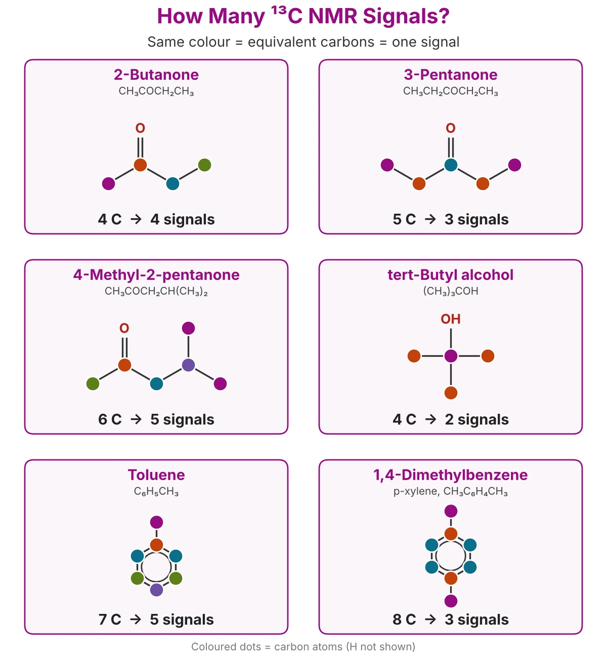
More Examples: How Many ¹³C NMR Signals?
Compare the Signals with the Molecular Formula
If the molecular formula has more carbons than signals, the molecule has symmetry. Example: C₅H₁₀O with only 3 signals must be 3-pentanone, not 2-pentanone, which gives 5.
Tip: two different carbons can, by chance, appear at almost the same δ and look like one line. So the number of signals is the smallest possible number of kinds of carbon; check doubtful cases with the chemical shift (Step 2) and DEPT (Step 3).
Step 2: Read the Chemical Shift: ¹³C NMR Chart and Table
The chemical shift (δ) of a signal tells you the kind of carbon. Read the δ of each signal, then match it with the ¹³C NMR chart (Figure 4) or the carbon NMR shifts table below. Carbons next to electronegative atoms (O, N) and sp² carbons (C=C, C=O) are deshielded and appear downfield (left); sp³ carbons in a hydrocarbon chain are shielded and appear upfield (right).
¹³C NMR Chart (Carbon NMR Shifts)
¹³C NMR Chemical Shift Table
The table gives the 13C NMR ranges with a measured example for each kind of carbon.
Simplified ¹³C NMR Ranges (Four Regions)
For a first look at an unknown spectrum, these four regions are enough:
Carbonyl Carbons: Ketone and Aldehyde vs Ester, Acid and Amide
Every C=O carbon is far downfield, but two groups can be told apart:
- Ketones and aldehydes: 190–220 ppm. Example: 4-methyl-2-pentanone, 209.0 ppm (Figure 1).
- Esters, acids and amides: 160–185 ppm. The O or N attached to the C=O carbon gives electron density to it by resonance, so the carbon is less deshielded. Example: the ester C=O of ethyl acetate, 171.4 ppm.
C=O above 190 ppm → aldehyde or ketone
C=O at 160–185 ppm → ester, acid or amide
Tip: the ranges overlap in places (for example, C=C alkene and aromatic carbons, or CH₃–C=O and CH), so use the chemical shift together with the number of signals (Step 1) and DEPT (Step 3). Exact values also change a little with the solvent and nearby groups.
Proton-Coupled and Proton-Decoupled ¹³C NMR Spectra
A ¹³C NMR spectrum can be recorded in two ways: proton-coupled, where each carbon signal is split by its hydrogens, or proton-decoupled, where the splitting is removed. Both are shown for 4-methyl-2-pentanone in Figure 5.
Proton-Coupled ¹³C NMR Spectrum
Each ¹³C nucleus is split by the hydrogens bonded directly to it (one-bond C–H coupling, ¹JC–H). The n + 1 rule applies, with n = the number of H on that carbon:
- Large coupling: ¹JC–H is about 125 Hz for sp³ carbons, 160 Hz for sp² carbons and up to about 250 Hz for sp carbons, much larger than H–H coupling (about 7 Hz).
- Result: the multiplets are wide (125 Hz = 1.25 ppm on a 100 MHz ¹³C scale), so neighbouring multiplets overlap (20–26 ppm in Figure 5) and the spectrum becomes crowded and weak.
Proton-Decoupled ¹³C NMR Spectrum
In a proton-decoupled spectrum (broadband decoupling, written ¹³C{¹H}), all the hydrogens are irradiated while the carbon spectrum is recorded. The C–H coupling disappears, so each kind of carbon gives one sharp line. This is the routine ¹³C NMR spectrum, and the one used in Steps 1 and 2.
- Stronger signals: each multiplet collapses into one line, and the nuclear Overhauser effect (NOE) from the irradiated hydrogens increases the carbon signal.
- No integration: the NOE and the relaxation time differ from carbon to carbon, so peak heights do not count carbons. Carbons with no hydrogen, such as the C=O at 209.0 ppm, are usually the smallest peaks.
- What is lost: the splitting that showed how many H each carbon carries.
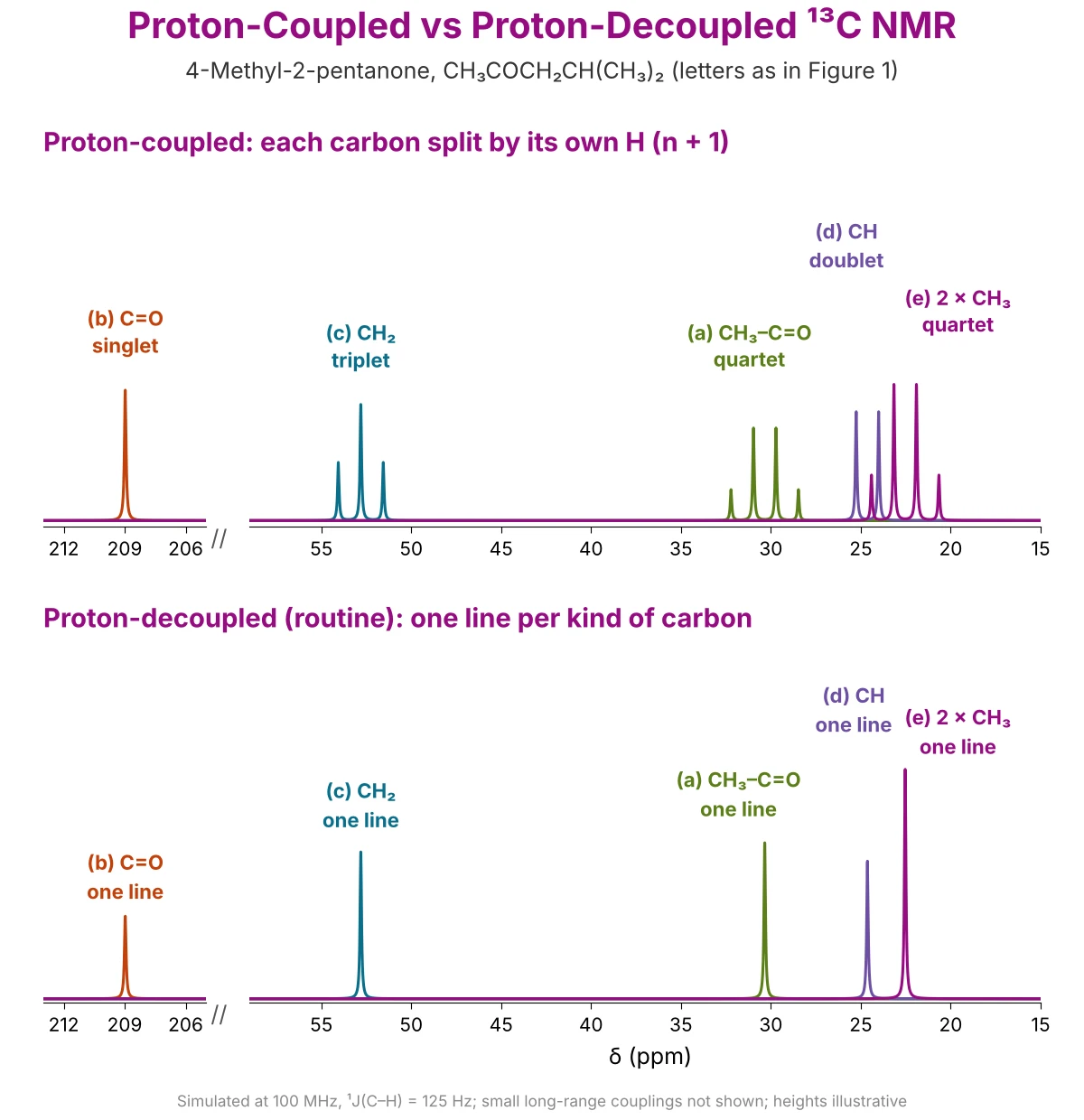
Proton-Coupled vs Proton-Decoupled ¹³C NMR
Best of both: the decoupled spectrum is clean but hides the number of H on each carbon; the coupled spectrum shows it but is crowded. DEPT gives this information while keeping one line per carbon; see Step 3: DEPT NMR.
Step 3: Identify Carbon Types with DEPT NMR (DEPT-90 and DEPT-135)
DEPT (distortionless enhancement by polarization transfer) tells you how many hydrogens each carbon carries: CH₃, CH₂, CH or C (no H). Magnetization is transferred from the ¹H nuclei to the ¹³C nuclei bonded to them, so only carbons with H give a DEPT signal. The last pulse angle, 90° or 135°, decides which carbons appear and whether they point up or down.
DEPT-90 → only CH (up)
DEPT-135 → CH₃ and CH up, CH₂ down
C with no H (C=O, quaternary C) → absent in both
How to Read DEPT Spectra
Run three spectra (the normal decoupled ¹³C, DEPT-135 and DEPT-90), then compare them signal by signal:
- CH: the peaks in DEPT-90.
- CH₂: the negative (downward) peaks in DEPT-135.
- CH₃: the positive DEPT-135 peaks that are not in DEPT-90.
- C with no H: the peaks in the decoupled spectrum that are missing from DEPT-135.

Proton-Decoupled Spectrum vs DEPT-135 vs DEPT-90: 4-Methyl-2-pentanone
All five signals appear in the decoupled spectrum (Figure 1). Reading them across the two DEPT spectra gives the type of every carbon:
Tips: add up the H found by DEPT and compare with the molecular formula; any missing H are on O or N (OH, NH), which DEPT does not show. The CDCl₃ solvent peak (77.16 ppm) is also absent in DEPT, because its carbon carries deuterium, not hydrogen.
Solvent Peaks in ¹³C NMR
¹³C NMR spectra are run in a deuterated solvent. Its hydrogens are replaced by deuterium, but its carbon atoms still give a ¹³C signal, so the solvent peak appears in every spectrum. It is not part of the sample: find it and leave it out when you count signals (Step 1). It is also often used as the reference instead of TMS.
¹³C NMR Solvent Peaks Table
Values from Fulmer et al. (2010). The ¹H column gives the residual solvent peak in ¹H NMR, for comparison with the ¹H NMR article.
Why the Solvent Peak Is a Triplet or a Septet
Deuterium (D) has a nuclear spin of 1, and it stays coupled to carbon in a proton-decoupled spectrum, because only ¹H is decoupled. A carbon bonded to n deuterium atoms gives 2n + 1 lines:
Number of lines = 2nI + 1 (I = 1 for D)
- CDCl₃: 1 D → 2(1)(1) + 1 = 3 lines, a 1 : 1 : 1 triplet at 77.16 ppm.
- CD₂Cl₂: 2 D → 5 lines (quintet).
- CD₃ groups (DMSO-d₆, acetone-d₆, CD₃OD, CD₃CN): 3 D → 7 lines (septet).
- Carbon with no D (C=O of acetone-d₆, C≡N of CD₃CN): one line.
DMSO Carbon NMR Peak (DMSO-d₆)
In DMSO-d₆, (CD₃)₂SO, the two CD₃ carbons are equivalent and give one septet at 39.52 ppm. It sits in the sp³ region (0–50 ppm), so check that a signal near 39–40 ppm is not the solvent before assigning it to a CH₂ or CH of the sample.
Tips: the solvent peak is absent in DEPT spectra (its carbon carries D, not H), which is a quick way to recognise it. Its exact position also changes slightly with temperature and concentration, so use the table values as a guide.
Worked Example: Identify an Unknown from Its ¹³C NMR and DEPT Spectra
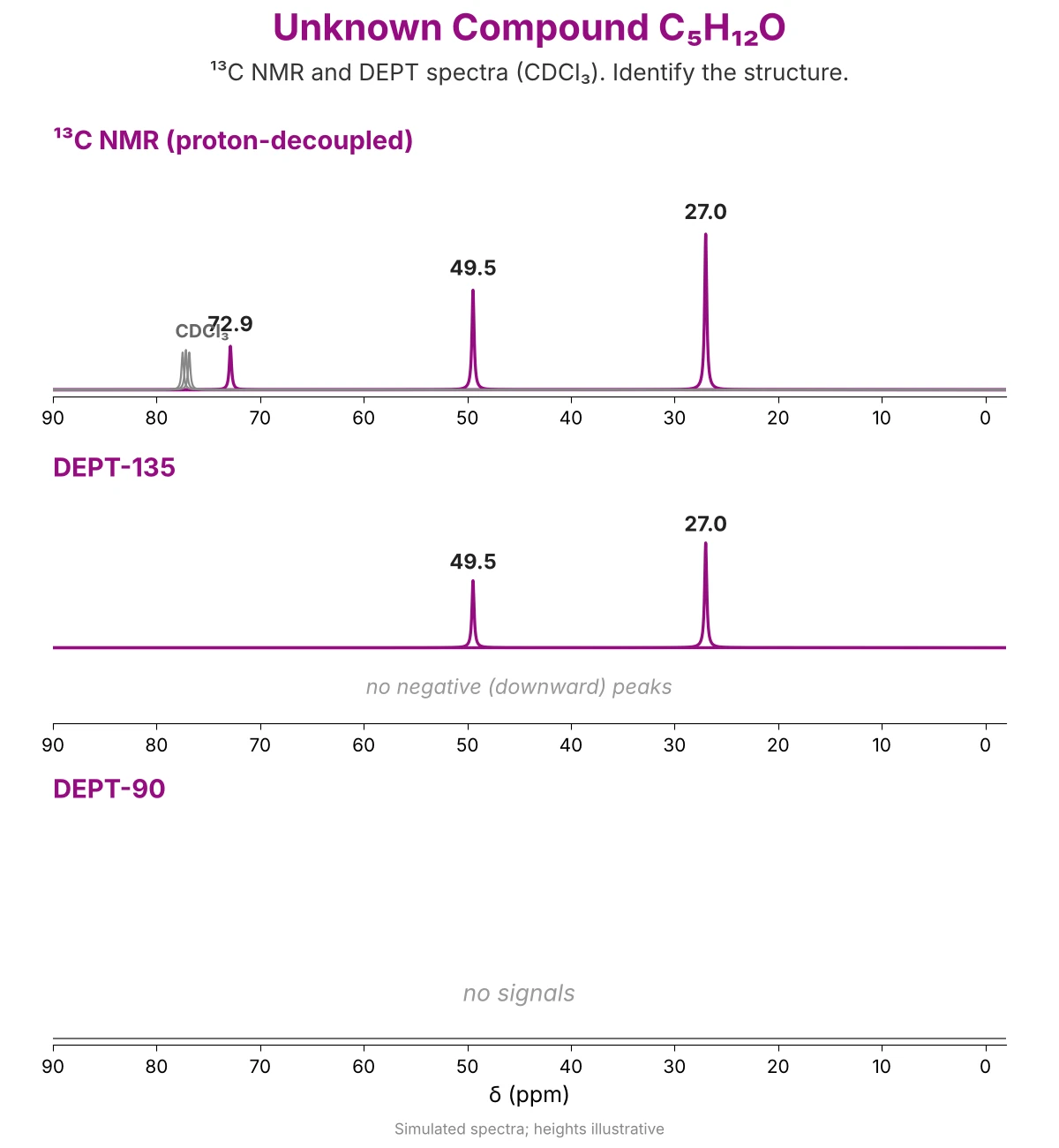
Try this before reading the answer. Use Steps 1–3 on the unknown spectra in Figure 7.
Solution, Step by Step
- Step 0: Use the molecular formula. C₅H₁₂O has DBE = (2 × 5 + 2 − 12) / 2 = 0: no ring, no C=C and no C=O. The one O must be in an alcohol or an ether.
- Step 1: Count the signals. Leave out the CDCl₃ triplet at 77.16 ppm. There are 3 signals for 5 carbons, so some carbons are equivalent: the molecule has symmetry.
- Step 2: Read the chemical shift. 72.9 and 49.5 ppm are in the C–O region (about 50–90 ppm), so two carbons are bonded to O. 27.0 ppm is an sp³ alkyl carbon.
- Step 3: Read the DEPT spectra. DEPT-90 shows nothing: no CH. DEPT-135 has no downward peak: no CH₂. 49.5 and 27.0 are up in DEPT-135 but absent in DEPT-90: both are CH₃. 72.9 is missing from DEPT-135: a carbon with no H, bonded to O.
- Step 4: Put the pieces together. Count the H: the 12 H of C₅H₁₂O must be on CH₃ groups only (no CH, no CH₂). The O can carry only one more group, so only one CH₃ (49.5, in the C–O region) is on O; the other three CH₃ (27.0, 9 H = three equivalent CH₃) must be on the carbon with no H (72.9). All 12 H are on carbon, so there is no OH: the compound is an ether, (CH₃)₃C–O–CH₃.
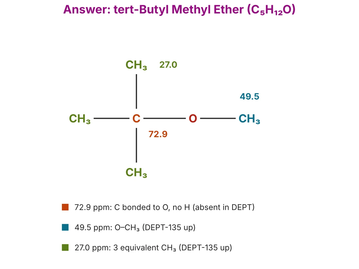
Check with ¹H NMR: tert-butyl methyl ether shows two singlets, 1.19 ppm (9H) and 3.22 ppm (3H), with no splitting, because no hydrogen has another hydrogen on the next carbon. An alcohol isomer such as 2-methyl-2-butanol, (CH₃)₂C(OH)CH₂CH₃, would instead show a CH₂ (downward in DEPT-135) and 4 ¹³C signals.
Using ¹H and ¹³C NMR Together
¹³C NMR and ¹H NMR answer different questions, so a structure is usually solved with C NMR and HNMR together: the ¹³C and DEPT spectra give the pieces (the kinds of carbon and their H), and the ¹H spectrum shows how the pieces are joined.
Which Spectrum Answers Which Question?
A Five-Step Plan for an Unknown
- 1. Formula and DBE. From the molecular formula, work out the degree of unsaturation (rings + π bonds).
- 2. ¹³C NMR. Count the kinds of carbon and read the δ regions: C=O (and which kind), C=C or aromatic, C–O.
- 3. DEPT. Label every carbon CH₃, CH₂, CH or C; add up the H and compare with the formula (missing H = OH or NH).
- 4. ¹H NMR. Use integration and splitting to join the pieces: which group sits next to which.
- 5. Check. Draw the structure and match every ¹³C and ¹H signal to it.
Example: 4-Methyl-2-pentanone
- ¹³C + DEPT give the pieces: a ketone C=O (209.0), one CH₂ (52.8), one CH (24.6) and two kinds of CH₃ (30.3 and 22.5).
- ¹H joins them: the CH₃ singlet at 2.12 ppm (3H) has no H neighbours and its δ fits CH₃–C=O; the CH₃ doublet at 0.92 ppm (6H) means two equivalent CH₃ on a CH, an isopropyl group; the CH₂ doublet at 2.30 ppm (2H) sits between the C=O and that CH.
- Result: CH₃–CO–CH₂–CH(CH₃)₂, with every signal matched.
Practice with both spectra: How to Read NMR Spectra (¹H NMR) covers integration and splitting; the 2-hexanone and ethyl acetate articles solve a structure from the mass, IR, ¹H and ¹³C NMR spectra together.
Common Mistakes in Reading ¹³C NMR Spectra
Most errors in ¹³C NMR interpretation come from reading it like a ¹H spectrum. Figure 9 sums up the six most common mistakes.
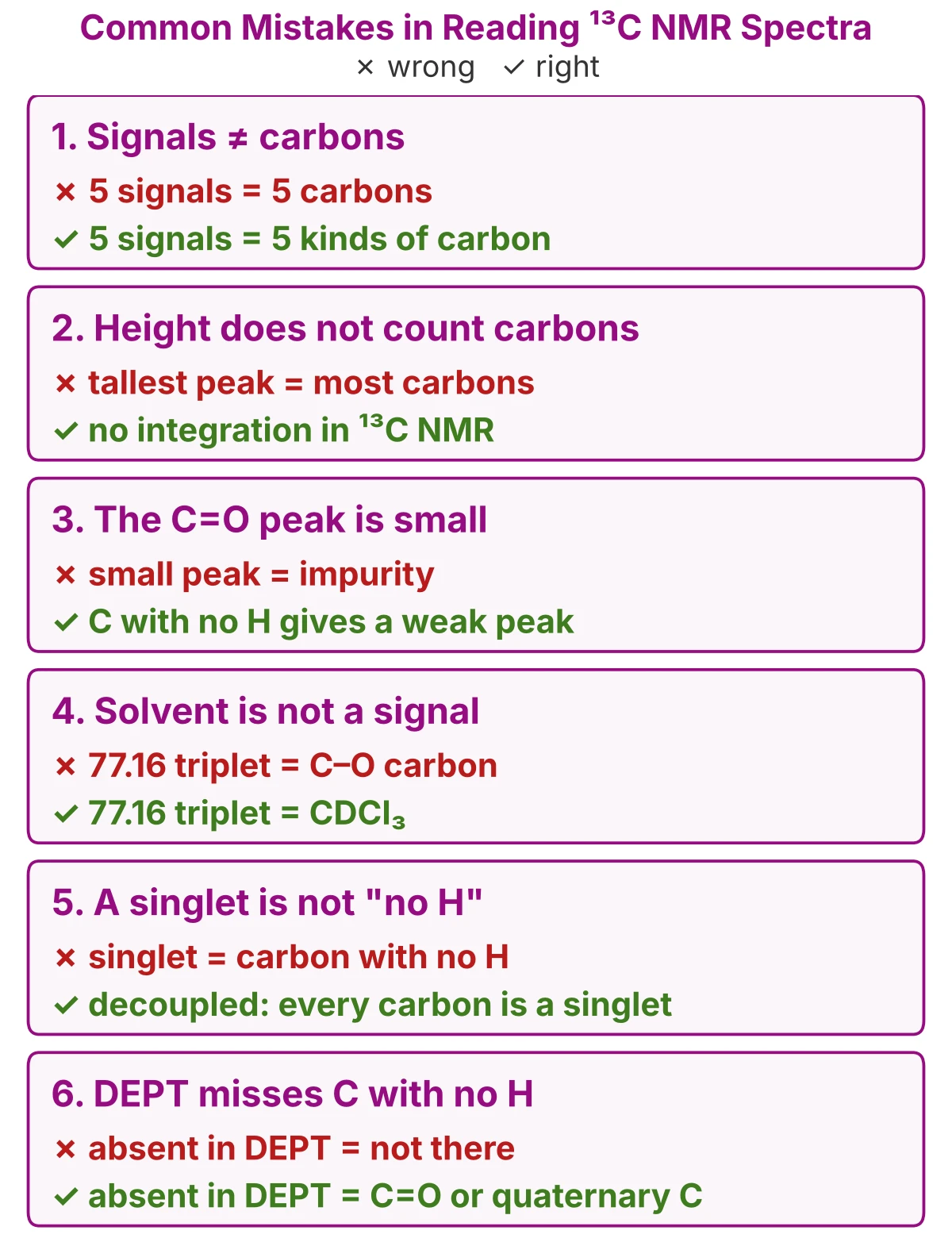
- Counting signals as carbons. Equivalent carbons give one signal, so 5 signals mean 5 kinds of carbon; compare with the molecular formula (Step 1).
- Using peak height to count carbons. In the routine decoupled spectrum, NOE and relaxation make peak sizes uneven, so ¹³C NMR is not integrated (proton-decoupled spectrum).
- Taking a small peak for an impurity. Carbons with no H, such as C=O and quaternary C, usually give the smallest peaks; a weak line near 160–220 ppm is often the C=O.
- Counting the solvent. The 1 : 1 : 1 triplet at 77.16 ppm is CDCl₃ and the septet at 39.52 ppm is DMSO-d₆; leave them out (solvent peaks).
- Reading a singlet as “no H”. In a proton-decoupled spectrum every carbon is a singlet, whatever its H; use DEPT to find the H (Step 3).
- Thinking a carbon missing from DEPT is not there. DEPT shows only carbons bonded to H; a peak in the ¹³C spectrum that is absent in DEPT-135 is a carbon with no H, such as C=O, C≡N or a quaternary carbon.
13C NMR: Practice MCQs with Answers
Twenty questions on reading a ¹³C NMR spectrum: questions 1–15 test each step, the last five test the ideas behind them. Pick an answer, then open it to check.
1. 13C NMR (carbon NMR), number of signals: how many signals does 3-pentanone, CH₃CH₂COCH₂CH₃, give in its C13 NMR spectrum?
- 2
- 3
- 4
- 5
- 6
Show answer
Answer: B. A mirror plane through the C=O makes the two ethyl groups equivalent, so 5 carbons give 3 signals: CH₃, CH₂ and C=O. (Step 1)
2. C NMR chart, carbon NMR shifts: where does the C=O carbon of a ketone appear?
- 0–50 ppm
- 50–90 ppm
- 115–140 ppm
- 160–185 ppm
- 200–220 ppm
Show answer
Answer: E. Ketone C=O carbons appear at 200–220 ppm; 160–185 ppm is the C=O of acids, esters and amides. (Step 2)
3. DEPT NMR, DEPT-135: which carbon gives a negative (downward) peak in a DEPT-135 spectrum?
- CH₂
- CH₃
- CH
- C=O
- Quaternary C
Show answer
Answer: A. In DEPT-135, CH₃ and CH point up, CH₂ points down, and carbons with no H are absent. (Step 3)
4. C-13 isotope, carbon-13 NMR: about what percentage of natural carbon is carbon-13?
- 0.01 %
- 1.1 %
- 12 %
- 50 %
- 98.9 %
Show answer
Answer: B. Carbon-13 is about 1.1 % of natural carbon; carbon-12 (98.9 %) has spin 0 and gives no NMR signal. (Why Carbon-13?)
5. CNMR solvent peak, C NMR spectrum in CDCl₃: where does the 1 : 1 : 1 triplet of the solvent appear?
- 0.00 ppm
- 7.26 ppm
- 39.52 ppm
- 77.16 ppm
- 128.06 ppm
Show answer
Answer: D. CDCl₃ gives a triplet at 77.16 ppm; 7.26 ppm is its residual ¹H peak and 39.52 ppm is DMSO-d₆. (Solvent peaks)
6. Carbon 13 NMR, DEPT-90: which carbons appear in a DEPT-90 spectrum?
- Only CH
- CH and CH₃
- Only CH₂
- All carbons
- Only carbons with no H
Show answer
Answer: A. DEPT-90 shows only CH carbons; CH₃, CH₂ and carbons with no H are absent. (Step 3)
7. 13C NMR table, ester C NMR: at what δ does the ester C=O carbon of ethyl acetate appear (CDCl₃)?
- 21.0 ppm
- 60.5 ppm
- 128.4 ppm
- 171.4 ppm
- 207.1 ppm
Show answer
Answer: D. The ester C=O of ethyl acetate is at 171.4 ppm; 60.5 is the O–CH₂, 21.0 the CH₃–C=O, 207.1 is acetone’s ketone C=O and 128.4 is benzene. (Step 2)
8. Proton-coupled 13C NMR spectrum: how does a CH₃ carbon appear when the spectrum is not proton-decoupled?
- Singlet
- Doublet
- Triplet
- Quartet
- Quintet
Show answer
Answer: D. A carbon is split by its own H (n + 1): CH₃ has 3 H, so it gives a quartet. (Proton-coupled spectrum)
9. Carbon-13 isotope: how many neutrons does carbon-13 have?
- 6
- 7
- 8
- 12
- 13
Show answer
Answer: B. Carbon-13 has 6 protons and 7 neutrons (6 + 7 = 13); carbon-12 has 6 neutrons. (Why Carbon-13?)
10. 13C NMR ranges, carbon NMR shifts: where does a carbon bonded to oxygen in an alcohol or ether appear?
- 0–20 ppm
- 20–40 ppm
- 50–90 ppm
- 100–150 ppm
- 190–220 ppm
Show answer
Answer: C. C–O carbons appear at about 50–90 ppm, for example ethanol CH₂O at 58.3 and diethyl ether at 65.9 ppm. (Step 2)
11. 13C NMR chart (C NMR chart, CNMR chart): in which range does a CH₃ carbon at the end of an alkyl chain appear?
- 10–16 ppm
- 50–90 ppm
- 115–140 ppm
- 160–185 ppm
- 200–220 ppm
Show answer
Answer: A. A chain-end CH₃ (R–CH₃) is among the most shielded carbons, at about 10–16 ppm. (Step 2)
12. Carbon NMR, C NMR table: what does the ¹³C NMR spectrum of benzene (C₆H₆) show?
- 1 signal at 77.16 ppm
- 1 signal at 128.4 ppm
- 3 signals at about 128 ppm
- 6 signals at 125–150 ppm
- 1 signal at 207 ppm
Show answer
Answer: B. All six benzene carbons are equivalent, so they give one signal, at 128.4 ppm in the aromatic region. (Step 1)
13. CNMR, C NMR spectrum: what does each line in a proton-decoupled C-13 NMR spectrum represent?
- One hydrogen
- One carbon atom
- One kind of carbon
- One bond
- One functional group
Show answer
Answer: C. Each line is one kind of carbon; equivalent carbons share one line, just as equivalent hydrogens do in ¹H NMR. (What Is a ¹³C NMR Spectrum?)
14. DEPT NMR, C NMR DEPT, carbon NMR: which DEPT result identifies a CH₃ carbon?
- Up in DEPT-90
- Down in DEPT-135
- Absent in both DEPT spectra
- Up in DEPT-135, absent in DEPT-90
- Up in both DEPT spectra
Show answer
Answer: D. CH₃ is positive in DEPT-135 but absent in DEPT-90; up in both is CH, down in DEPT-135 is CH₂, absent in both is a carbon with no H. (Step 3)
15. 13C NMR, C13 NMR, carbon NMR shifts: which signal of 4-methyl-2-pentanone is farthest downfield?
- 22.5 ppm (2 × CH₃)
- 24.6 ppm (CH)
- 30.3 ppm (CH₃–C=O)
- 52.8 ppm (CH₂)
- 209.0 ppm (C=O)
Show answer
Answer: E. The ketone C=O carbon is the most deshielded, at 209.0 ppm, far left of the sp³ carbons. (Step 2)
16. Why is a routine ¹³C NMR spectrum not integrated to count carbons?
- There is no TMS signal
- There are too few signals
- The NOE and relaxation differ from carbon to carbon
- The solvent covers the peaks
- Carbon-13 has no nuclear spin
Show answer
Answer: C. In a proton-decoupled spectrum, the NOE and relaxation times differ between carbons, so peak areas do not match the number of carbons. (Proton-decoupled spectrum)
17. A signal at 170 ppm appears in the decoupled ¹³C spectrum but in neither DEPT-135 nor DEPT-90. What is it?
- A carbon with no H, such as an ester C=O
- A CH₂
- A CH₃
- An impurity
- A CH
Show answer
Answer: A. Carbons with no H do not appear in DEPT; at 170 ppm this is a C=O of an ester, acid or amide. (Step 3)
18. Why are ¹³C NMR signals not split by neighbouring carbons?
- Carbon-12 has spin ½
- Decoupling removes C–C coupling
- Carbon has no nuclear spin
- DEPT removes the splitting
- ¹³C atoms are rare, so two are almost never next to each other
Show answer
Answer: E. Only 1.1 % of carbons are ¹³C, so two ¹³C atoms are almost never bonded together and C–C splitting is not seen. (Why Carbon-13?)
19. An unknown C₄H₈O shows four ¹³C signals at about 209, 37, 29 and 8 ppm. In DEPT-135, 37 points down and 29 and 8 point up; 209 is absent. What is the compound?
- Butanal
- 2-Methylpropanal
- 2-Butanone
- Tetrahydrofuran
- Cyclobutanol
Show answer
Answer: C. 209 ppm with no H is a ketone C=O; one CH₂ (37) and two CH₃ (29, 8) give CH₃COCH₂CH₃, 2-butanone, a methyl ketone that also gives a positive iodoform test. An aldehyde would show a CHO carbon (a CH) at 190–200 ppm in DEPT-90. (Worked example)
20. Why does CDCl₃ give a triplet in a proton-decoupled ¹³C NMR spectrum?
- Coupling to the three Cl atoms
- Coupling to hydrogen
- It is a DEPT effect
- It is caused by the NOE
- Coupling to one deuterium (spin 1) gives 2nI + 1 = 3 lines
Show answer
Answer: E. Only ¹H is decoupled; the carbon stays coupled to one D (I = 1), giving 2(1)(1) + 1 = 3 lines. (Solvent peaks)
13C NMR: Frequently Asked Questions
How do you interpret a 13C NMR spectrum (how to read carbon NMR)?
Count the signals to find the kinds of carbon, read each chemical shift against a ¹³C NMR chart to find the type of carbon, then use DEPT to label each one CH₃, CH₂, CH or C. Compare the result with the molecular formula and the ¹H NMR spectrum.
What is the basic principle of 13C NMR spectroscopy?
The ¹³C nucleus has a spin of ½, so in a strong magnetic field it absorbs radio waves at a frequency that depends on its chemical environment. Each kind of carbon therefore appears at its own chemical shift (δ, ppm from TMS).
What is 13C NMR used for?
Carbon NMR shows the carbon skeleton of a molecule: how many kinds of carbon it has, which functional groups are present (C=O, C=C, C–O) and, with DEPT, how many H each carbon carries. It is used with ¹H NMR, IR and mass spectrometry to identify organic compounds.
Why is C-13 used in NMR instead of C-12?
Carbon-12, the main isotope (98.9 %), has a nuclear spin of zero and gives no NMR signal. Carbon-13 (about 1.1 %) has a spin of ½, so it is the only carbon isotope that NMR can detect.
Why is C-13 NMR less sensitive than 1H NMR?
Only 1.1 % of carbon atoms are ¹³C, and the ¹³C nucleus is a weaker magnet than ¹H (it resonates at about one-fourth the frequency), so its signal is about 6000 times weaker. More sample and many scans are needed.
How many signals are in a 13C NMR spectrum (how to count peaks in C NMR)?
Count one signal for each kind of carbon: equivalent carbons, related by symmetry, give one signal. Leave out TMS and the solvent peak, for example the CDCl₃ triplet at 77.16 ppm.
What is the range of C-13 NMR (13C NMR chart)?
¹³C chemical shifts cover about 0–220 ppm: sp³ carbons at 0–50, C–O at 50–90, C=C and aromatic carbons at 100–150, and C=O at 160–220 ppm. This wide range means ¹³C signals rarely overlap.
Is 13C NMR in ppm, and how is the chemical shift calculated?
Yes. The chemical shift is δ (ppm) = (frequency of the signal − frequency of TMS, in Hz) ÷ spectrometer frequency (MHz), so the same compound gives the same δ on any instrument.
Does 13C NMR have integration?
Not in the routine spectrum. Because of the nuclear Overhauser effect and different relaxation times, peak areas in a proton-decoupled ¹³C spectrum do not match the number of carbons, so peaks are counted, not integrated.
What are the two main types of NMR spectroscopy?
¹H NMR (proton NMR), which studies hydrogens, and ¹³C NMR (carbon NMR), which studies carbons. Together they give the hydrogen and carbon frameworks of a molecule.
What is the N-1 rule in NMR?
There is no N − 1 rule; the rule is n + 1. In ¹H NMR, n is the number of H on neighbouring carbons; in a proton-coupled ¹³C spectrum, n is the number of H bonded to the carbon itself, so CH₃ gives a quartet.
What is DEPT in 13C NMR (DEPT NMR)?
DEPT (distortionless enhancement by polarization transfer) shows which carbons carry H: DEPT-90 shows only CH, and DEPT-135 shows CH₃ and CH up and CH₂ down. Carbons with no H are absent from both.
How is a 13C NMR spectrum processed?
The signal recorded by the instrument (the FID) is converted into a spectrum by a Fourier transform; the spectrum is then phased, its baseline corrected and its δ scale referenced to TMS (0.00 ppm) or the solvent (CDCl₃, 77.16 ppm).
What are the limitations of 13C NMR?
It is slow and needs more sample because of the low abundance of ¹³C, peak areas cannot be used to count carbons, and the routine spectrum does not show how many H each carbon carries unless DEPT is run.
How many protons, neutrons and electrons are in carbon-13?
Carbon-13 has 6 protons and 7 neutrons in its nucleus, and a neutral atom has 6 electrons; its atomic number is 6 and its mass number is 13.
Is C-13 radioactive?
No. Carbon-13 is a stable isotope; the radioactive isotope of carbon is carbon-14, which is used in radiocarbon dating.
What is a CNMR predictor?
A CNMR predictor is software that estimates the ¹³C chemical shifts of a structure from databases and additivity rules. It is useful for checking an assignment, but measured values can differ by a few ppm.
13C NMR: Key Terms
¹³C NMR (carbon NMR): nuclear magnetic resonance of carbon-13 nuclei; it shows the kinds of carbon in a molecule (the carbon skeleton). Example: 4-methyl-2-pentanone gives five ¹³C signals (Figure 1).
Carbon-13 (¹³C, C-13 isotope): the stable carbon isotope with 6 protons and 7 neutrons, about 1.1 % of natural carbon, with nuclear spin ½. Example: only about 1 carbon atom in 90 is ¹³C, so ¹³C NMR signals are weak.
Chemical shift (δ) in ¹³C NMR: the distance of a carbon signal from the TMS signal (0.00 ppm), measured in ppm; it shows the type of carbon (range about 0–220 ppm). Example: the ketone C=O of acetone is at 207.1 ppm.
Proton-decoupled (broadband-decoupled) spectrum: the routine ¹³C spectrum, recorded while all ¹H are irradiated, so each kind of carbon gives one line. Example: 4-methyl-2-pentanone shows five single lines.
Coupling constant in ¹³C NMR (¹JC–H): the splitting between a ¹³C and the H bonded to it, in Hz. Example: about 125 Hz for an sp³ C–H.
DEPT NMR: distortionless enhancement by polarization transfer: a set of ¹³C experiments that shows how many H each carbon carries. Example: in DEPT-135, CH₃ and CH point up and CH₂ points down; DEPT-90 shows only CH; C=O is absent from both.
Solvent peak (CDCl₃ NMR peak): the ¹³C signal of the deuterated solvent; it is not part of the sample. Example: CDCl₃ gives a 1 : 1 : 1 triplet at 77.16 ppm.
Downfield vs upfield: downfield is the left of the spectrum (larger δ, deshielded carbon); upfield is the right (smaller δ, shielded carbon). Example: the C=O at 209.0 ppm is far downfield of the CH₃ at 22.5 ppm.
Learn More: NMR, IR and Mass Spectra Worked Examples
Read: ¹H NMR and worked examples with ¹³C NMR
How to Read NMR Spectra (¹H NMR) 2-Hexanone Spectra (Mass, IR, ¹³C and ¹H NMR) Ethyl Acetate Spectra and Structure Elucidation
Read: other spectroscopy
IR Spectrum Interpretation Mass Spectrum Interpretation What is Mass Spectrometry? Base Peak and Molecular Ion Peak
Try: free tools
IR Peak Identifier IR Spectrum Comparison Tool Mass Spectrum Fragment Identifier Molecular Mass Calculator
Identify the same compounds by chemical tests on chemistrySH.com
Identification of Acetone Identification of Benzaldehyde Iodoform Test for Methyl Ketones chemistrySH.com
Sources and Further Reading
[1] Bruice, P. Y. (2016). Organic chemistry (8th ed.). Pearson.
[2] Gottlieb, H. E., Kotlyar, V., & Nudelman, A. (1997). NMR chemical shifts of common laboratory solvents as trace impurities. The Journal of Organic Chemistry, 62(21), 7512–7515. https://doi.org/10.1021/jo971176v
[3] Fulmer, G. R., Miller, A. J. M., Sherden, N. H., Gottlieb, H. E., Nudelman, A., Stoltz, B. M., Bercaw, J. E., & Goldberg, K. I. (2010). NMR chemical shifts of trace impurities: Common laboratory solvents, organics, and gases in deuterated solvents relevant to the organometallic chemist. Organometallics, 29(9), 2176–2179. https://doi.org/10.1021/om100106e
[4] Babij, N. R., McCusker, E. O., Whiteker, G. T., Canturk, B., Choy, N., Creemer, L. C., De Amicis, C. V., Hewlett, N. M., Johnson, P. L., Knobelsdorf, J. A., Li, F., Lorsbach, B. A., Nugent, B. M., Ryan, S. J., Smith, M. R., & Yang, Q. (2016). NMR chemical shifts of trace impurities: Industrially preferred solvents used in process and green chemistry. Organic Process Research & Development, 20(3), 661–667. https://doi.org/10.1021/acs.oprd.5b00417